Magnetic resonance imaging (MRI)
What is MRI?
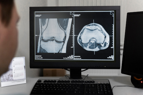
Magnetic resonance imaging (MRI) is a safe and informative diagnostic method based on the phenomenon of magnetic resonance, without the use of X-rays. It allows you to obtain clear images of internal organs, joints and soft tissues without the use of X-rays.
At the K+31 clinic, MRI is performed on expert-class equipment with high scanning speed, comfortable conditions for the patient and prompt delivery of results.
Branch K+31 on Lobachevsky:
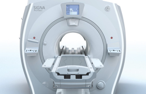
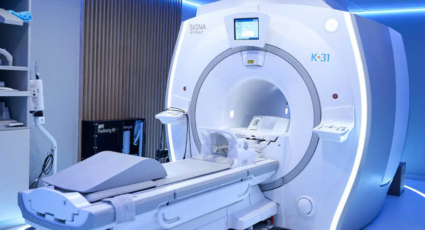
GE SIGNA™ Architect 3.0 Tesla
- Expert-level device with a magnetic field of 3 Tesla
- Ultra-high image resolution
- Fast scanning and reduced time the patient spends in the device
- Wide tunnel (70 cm) - comfort even for patients with claustrophobia
Branch K+31 West:
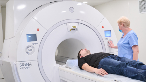
GE SIGNA™ Voyager 1.5 Tesla
- Universal device of the new generation
- Increased quality of soft tissue images
- Innovative system of accelerated scanning
Branch K+31 Petrovsky Gates:
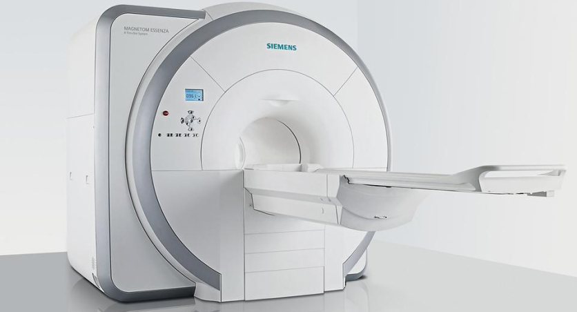
SIEMENS MAGNETOM Essenza 1.5 Tesla
- Reliable German-made device
- Excellent image quality with standard protocols
- The tunnel diameter is 60 cm - compact and convenient for patients with limited mobility
- More accurate diagnosis due to high detail
- Reduced scanning time - less time for research
- Ability to conduct research of any complexity: from standard to specialized protocols
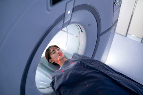
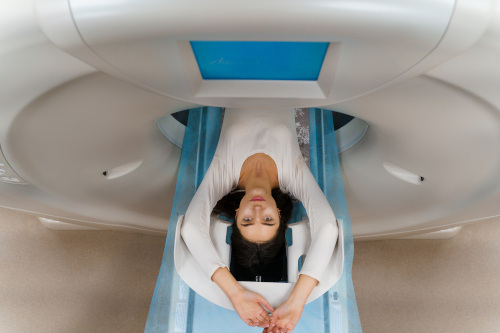
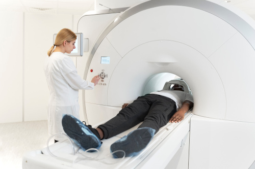
MRI is a completely painless procedure. The patient is placed on a retractable couch that moves inside the device. It is important to remain still during the examination - this allows you to obtain the most accurate and high-quality images. Scanning time varies depending on the area and purpose of the examination. If necessary (claustrophobia, children, elderly patients), individual conditions are possible.
In most cases, special preparation is not required. However, when examining certain areas, there are exceptions, including a number of absolute and relative contraindications. Read more at the links below:
Popular services
MRI under anesthesia
The specificity of MRI is that the patient must remain motionless for 20-90 minutes during the entire scanning period. Sometimes, due to exacerbation of claustrophobia, noise inside the coil or for other reasons, it is advisable to perform tomography under anesthesia. This procedure has its own characteristics.
This type of diagnostics with anesthesia is used in the following situations:
- Mental disorders, due to which the patient is unable to behave adequately
- Fear of closed spaces (claustrophobia), a tendency to panic attacks
- Pain, as a result of which the patient cannot remain still
- Hyperkinesis, neurological disorders that provoke involuntary muscle movements
Anesthesia always carries certain risks, especially for patients with heart disease, lung disease or drug allergies. Side effects such as nausea, dizziness or weakness after anesthesia are possible. The patient may feel drowsy or weak after anesthesia, so he is advised to rest for several hours. The MRI results are analyzed by a doctor, and based on them, a diagnosis is made or treatment is adjusted. If you or a loved one is going to have an MRI under anesthesia, it is important to discuss all the details with your doctor to minimize risks and ensure a comfortable procedure.

Appoint to MRI

Our doctors


This award is given to clinics with the highest ratings according to user ratings, a large number of requests from this site, and in the absence of critical violations.

This award is given to clinics with the highest ratings according to user ratings. It means that the place is known, loved, and definitely worth visiting.

The ProDoctors portal collected 500 thousand reviews, compiled a rating of doctors based on them and awarded the best. We are proud that our doctors are among those awarded.
Price


Answers to popular questions
What implants can be used for MRI?
With ceramic-metal dentures, crowns. They are not considered as implants. Also, MRI results do not depend on the presence of an intrauterine device. Any metal or ferromagnetic implants are questionable. This also applies to cochlear prostheses in the inner ear. In these cases, it is necessary to provide the doctor with an implant passport.
Is it possible to do an MRI during pregnancy and how does MRI affect the fetus?
Among the contraindications for MRI are the first 3 months of pregnancy, when the fetal organs are laid down. During this period, the procedure can be carried out by pregnant women, usually only in case of urgent need and only in the direction of a doctor. The device does not emit X-rays, as with computed tomography or X-rays. The magnetic field with which MRI works does not affect the development of the fetus in any way, and for 30 years of using the technology, negative cases have not been identified. Otherwise, the MRI method is the safest procedure and is used to diagnose the health of both the mother and the fetus. The device is able to detect a variety of malformations, diseases and collect all the information about the development of the fetus.
Does MRI affect the body while breastfeeding?
The electromagnetic waves and the magnetic field generated during the study do not affect the body of the nursing mothers and do not cause any changes in the composition of milk. After the MRI, you can immediately start feeding.
What is an MRI procedure?
The assistant helps the patient to sit on the table of the device. One of the conditions is the fixation of the examined area of the body to ensure immobility. For this, soft belts and rollers are used. The patient does not experience any discomfort he can blink, swallow, and talk to a doctor. The part of the body to be examined is located in the coil of the apparatus, emitting and receiving reflected magnetic waves.
Examination of one organ or part of the body takes 30-40 minutes, however, if it is a complex case, more time will be needed. The process itself is a series of shots that last for a few minutes each. During the operation of the device, you can not move, in the intervals a small movement is allowed, but the examined part of the body must remain motionless. Otherwise, the next series of shots will not match the previous one. To take a series of shots, the doctor will ask you to hold your breath for 10-15 seconds.
How is an MRI with contrast performed?
In some cases, only in the direction of a doctor, the procedure involves the use of a contrast agent. This method is used for suspected oncology, oncological processes and for the early detection of metastases that require immediate treatment. A special substance is first injected into a vein in the patient's arm through a catheter. It is an absolutely safe solution, which for some time makes the vessels of the organ under study (for example, the brain) visible to the MRI machine. It does not cause allergic and adverse reactions. During the day, the substance is completely excreted from the body in a natural way.
How does an MRI feel?
The procedure is completely painless. The only thing that can cause a feeling of discomfort is the need to be on time. studies alone in the office. But the doctor will be in the control room and talk to the patient on the speakerphone. Patients with a fear of confined spaces also have nothing to fear. Our apparatus leaves enough open space. In addition, the Siemens tomograph is characterized by an extremely low noise level during operation. If an MRI study is performed with a contrast agent, then when it is administered, the patient may feel cool, a slight rush blood, associated with the entry of a substance into the vessels. The sensations last no more than a couple of minutes.
Do I need to prepare for an MRI?
No, no special preparation is required. But the doctor needs to know about your past illnesses, recent injuries, surgeries, etc. If you are wearing metal jewelry or other metal items, you will need to remove them. If the doctor did not limit the intake of drugs, food before the MRI, no changes does not occur in the usual daily routine.
MRI at K+31
K+31 presents MRI machines in each clinic.
Siemens Magnetom Essenza 1.5 Tesla in K+31 Petrovsky Gates.
SIGNATM Architect AIRTM Edition 3 Tesla at K+31 Lobachevsky str.
GE Signa Voyager 1.5 Tesla in K+31 West.
MRI can be done with contrast (in the diagnosis of tumors and small anatomical structures) and without contrast, in a state of sleep or without anesthesia:
- Thyroid
- Liver
- Gallbladder and ducts
- Pancreas
- Kidney
- Spleens
- Joints
- Mammary glands
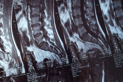
- Spinal Cord
- Arteries and veins of the head, neck, abdominal region
- Pelvic organs
- Soft tissues
- Lumbar
- Etc
Make an appointment at the medical center for an MRI or a doctor by phone: +7 (495) 162-66-97.
Conducting an MRI study
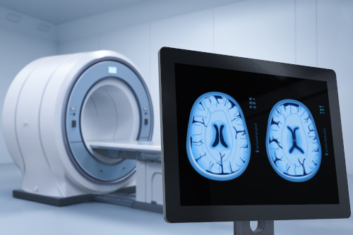
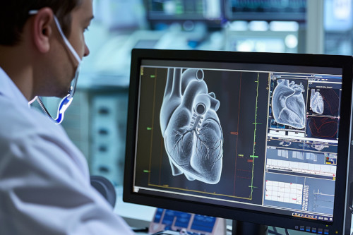
MRI is most often prescribed (in combination with other data from laboratory and instrumental studies), to clarify or establish a diagnosis, identify vascular anomalies, for early diagnosis of diseases brain, intervertebral discs, with multiple sclerosis, with suspected neoplasms (tumors), determining the pathology of internal organs, to assess surgical intervention and the consequences of injuries.
On average, an MRI examination lasts from 15 minutes to 1 hour, when using contrast, the time increases by 15 minutes.
Duration of MRI diagnostics:
- MRI of the knee joints – 20 minutes.
- MRI of the head, brain and MRI of neck vessels – 15 minutes.
- MRI of the mammary glands – 30 minutes.
- MRI of the abdominal cavity – 40-45 minutes.
- MRI of the lumbosacral spine - 15 minutes.
- MRI of the thoracic spine - 15 minutes.
- MRI of the cervical spine - 15 minutes.
- MRI of the pelvic cavity – 40 minutes.
- MRI of the bladder - 35 minutes.
During the examination, you must remain completely still. This should be taken into account in patients with severe pain. If, due to severe pain, it is not possible to remain in a forced position for a long time, then before the study it is necessary to perform seek pain relief from your doctor or come for an examination after relief of acute pain. If necessary, MRI is performed under sedation with the assistance of an anesthesiologist.
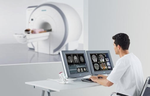
After MRI examination
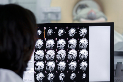
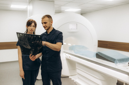
A written conclusion and a computer disk with the obtained images are issued to the patient on the day of the examination. In the pictures, areas of healthy tissues differ from those in which any changes occur, but interpret the images and give based on them, only a specialist can provide certain recommendations for further actions. After examination, based on the results of the scan, the doctor may recommend to consult by a neurologist, traumatologist, surgeon, urologist, gynecologist, etc. The final diagnosis is made your doctor and prescribes the appropriate treatment according to him.
Contraindications for MRI
MRI is not always a suitable diagnostic method, especially if there are certain contraindications. There are absolute guidelines for performing MRI. and relative contraindications. If the patient has absolute contraindications, the study cannot be performed, if there are relative contraindications, the question of conducting the study is decided individually with the MRI physician performing the examination.
Absolute contraindications to MRI:
- Presence of a pacemaker
- Insulin pumps
- Cerebral vessel clips after surgical treatment
- Large metal implants and fragments in the body
- Presence of metal shavings and splinters in the eye area
- Middle ear implants (fixed hearing aids)
- Weight limit - no more than 130 kg
Relative contraindications to MRI:
- Heart valve prostheses
- Pins
- Neurostimulants
- Claustrophobia (fear of enclosed spaces)
- Decompensated heart failure
- Hemostatic clips (except for cerebral vessels)
- For women - first trimester of pregnancy (first 13 weeks)
- Renal failure (on MR examination with contrast)
- Having an inner ear prosthesis
- Presence of tattoos, especially in the area being examined
- The need for cardiac monitoring
Reviews


How to find us

Application “Personal Account K+31”


