MRI under anesthesia (sedation)
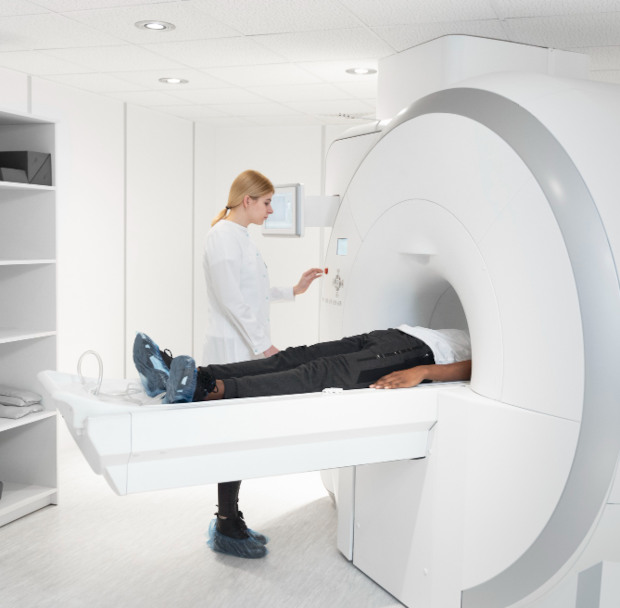
It's important to remain still during an MRI. This isn't always easy for an adult, and for a child or a patient with pain, anxiety, or claustrophobia, it can be nearly impossible. If a person moves, the images become less clear, and the examination must be repeated.
MRI under anesthesia helps ensure a calm, stress-free examination with the required image quality.

specialists

equipment

treatment


How is sedation different from general anesthesia?
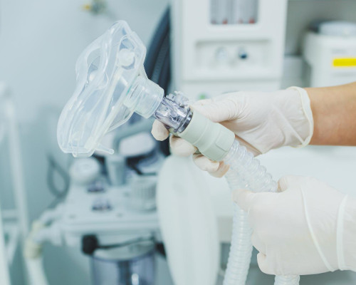
Sedation is usually easier to tolerate because it is gentler than general anesthesia. The patient falls asleep, is unaffected by the noise of the CT scanner, and lies quietly during the scan. Breathing often remains spontaneous, and the doctor constantly monitors the pulse, blood pressure, and blood oxygen levels.
General anesthesia is chosen in more complex cases when deep immobility is required and sedation is no longer sufficient. The final decision is made by the anesthesiologist after an examination.
Why is this important for a high-quality and accurate diagnosis?
Even slight movement of the head, back, or limb can interfere with a series of images. This is especially critical when examining the brain, blood vessels, spine, or postoperative area.
Therefore, anesthesia during MRI helps the doctor obtain data that can be used to make further decisions.
Indications for MRI under anesthesia
Indications are determined based on the patient's condition, not simply the desire to undergo a sleep MRI. The doctor considers age, pain, anxiety, neurological symptoms, scan duration, and the need for contrast. If a standard procedure has already failed, sleep MRI helps avoid a repeat attempt.
Reasons to discuss support in advance:
- Severe claustrophobia
- Tremor or hyperkinesis
- Pain when lying down
- Age under 7 years
- Long-term examination
After the assessment, the doctor chooses sedation, deeper sleep, or general anesthesia for the MRI. Sedatives should not be taken without a doctor's approval. Medications taken without a doctor's approval can alter breathing, blood pressure, and the response to anesthesia.
MRI for a child under anesthesia: features and necessity
It's difficult for a child to lie still for 20-40 minutes, not speak, and not react to noise. Therefore, performing an MRI on a child under anesthesia is sometimes the only way to obtain high-quality images the first time. Parents should inform the doctor about medications, allergies, fever, cough, and runny nose.
If children have recently been ill, the examination may be postponed until the airway has recovered. An MRI for a child under anesthesia is performed after the anesthesiologist has cleared the child, the dose has been calculated, laboratory tests have been reviewed, and the recovery room has been prepared. After the examination, the child remains under observation until their condition is stable.
Indications for adults: claustrophobia, hyperkinesis, pain syndrome
In adults, a common cause is a panic reaction to a confined space. The person understands the need for diagnosis, but cannot remain motionless inside the machine. In such cases, an MRI under sedation helps complete the examination without disruption.
Patient pain can make it difficult to maintain a supine position. Tremors, hyperkinesis, and stroke sequelae can cause involuntary movements. In these cases, anesthesia during MRI allows for obtaining images without re-scanning.
Emergency and planned cases of examination
A routine procedure is scheduled after a consultation, once it is clear why the standard format is not suitable. Urgent decisions are made in the following cases:
- In case of injury
- Seizures
- Speech impairment
- Weakness in the limbs
- Suspected space-occupying lesion
If a brain scan is required, immobility is especially important for assessing small structures.
For urgent indications, preparation is quicker, but a safety check is not skipped. The doctor clarifies the patient's history of implants, allergies, ongoing medications, chronic diseases, and previous examination results. This is necessary to avoid wasting time and increasing risks to the patient.
If a patient is unable to undergo the examination as usual, the doctor may suggest a sleep MRI.
General information about the procedure
Preparation for an MRI
Preparation for an MRI with anesthesia begins before the day of the appointment. The patient undergoes an examination, receives a list of tests, and discusses ongoing medications. This allows the doctor to select medications and reduce the risk of complications.
Consultation with an anesthesiologist and necessary tests
An appointment is necessary before the examination, not on the day of the procedure in a rush. The doctor will review chronic illnesses, allergies, reactions to previous anesthesia, medication use, and respiratory characteristics. The following are commonly prescribed:
- Complete blood count
- ECG
- Sometimes a blood chemistry test
- Additional findings as indicated
Fasting and medication guidelines before the examination
Before sedation or anesthesia, it is important to fast and drink no food or liquid. The doctor will determine the exact time in advance, taking into account the patient's age, health status, and the chosen anesthesia method.
Preparation for an MRI also includes checking your regular medications. You cannot stop taking medications on your own: some should be continued, while others can only be adjusted with your doctor's approval.
What to bring to the clinic on the day of the examination
On the day of the examination, you should arrive early so the team can calmly review your documents and prepare you. If contrast is planned, the doctor will check your tolerance to the medications and determine your renal function, as indicated. Bring your basic documents and medical information.
- Passport and referral
- Test results
- Extracts and images
- List of medications
- Metal-free clothing
After the check, the patient begins the examination and preparation. Children can wear their usual clothing; Only non-metallic items are allowed into the office.

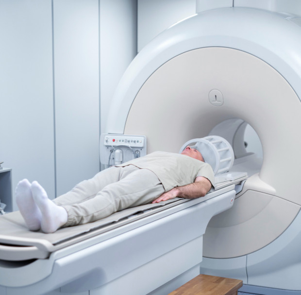
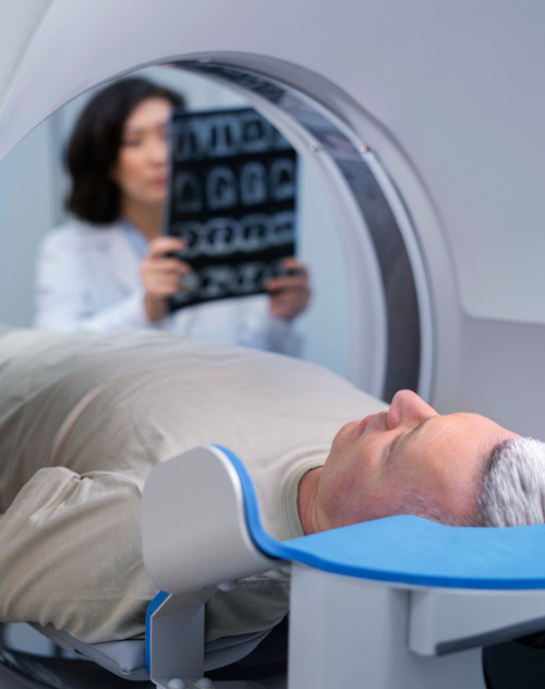
Modern high- and ultra-high-field CT scanner
The quality of diagnostics depends on the protocol, the radiologist's experience, and the equipment's capabilities. A modern CT scanner helps obtain detailed images, and patient immobility improves the accuracy of the results. This is especially important for long-term programs.
Comfortable recovery rooms and attentive support
Recovery from an MRI under anesthesia is monitored. The doctor and nurse ensure the patient is breathing normally, responding to treatment, and not complaining of severe nausea, pain, or dizziness.
Once the patient's condition has stabilized, they provide clear instructions for home care. Drowsiness upon awakening is common in children and usually resolves gradually. If unusual symptoms appear, it's best not to wait—discuss them with your doctor.
Before choosing a format, it's helpful to compare options. The chart helps you understand the differences but is not a substitute for a physical examination.
|
Signature |
MRI without anesthesia |
Drug-induced sedation |
General anesthesia |
|---|---|---|---|
|
Consciousness |
Full |
Sleep, relaxation |
Deep sleep |
|
Breathing |
Independent |
Independent |
Sometimes support is needed |
|
Indications |
No fear or pain |
Age, fear, discomfort |
Complex cases |
|
Recovery |
Not necessary |
1–2 hours |
From 2 hours |
The choice depends on the diagnosis, age, breathing, heart, weight, and the scope of the examination. The doctor selects the format to obtain accurate images and not increase unnecessary risks.
Benefits of MRI under sedation at K+31
The K+31 Clinic combines radiology and anesthesiology services in a single process. Each step is explained to the patient, and the depth of anesthesia is determined after the examination.
This approach is especially important if anesthesia is required for an MRI scan for a child, an elderly person, or a patient with chronic diseases.
Experienced Team of Anesthesiologists and Intensivists
An anesthesiologist and intensivist provides care to the patient before, during, and after the scan. They select medications based on age, weight, diagnosis, and scan duration. When performing anesthesia for a pediatric patient, this assessment is especially important because the response to medications depends on many factors.
Doctor's quote: "In my experience, the fear of anesthesia is often greater than the risks of the procedure itself. At K+31, we use modern, short-acting medications. Our goal is to ensure patient safety and comfort at every stage, from consultation to discharge."
Frequently Asked Questions
What is sedation for MRI in simple terms?
Sedation is controlled sleep or relaxation using medications. The patient remains motionless and experiences no significant discomfort. MRIs under sedation are performed under the supervision of an anesthesiologist.
Is anesthesia dangerous for a child during an MRI?
Any anesthesia has risks, but examination and monitoring during the procedure reduce them. Medications are selected based on age, weight, and health status. Therefore, MRIs are performed on children under general anesthesia only after clearance from an anesthesiologist.
What tests are needed before the study?
Most often, a complete blood count and ECG are required, sometimes a biochemical analysis and additional reports. The list depends on the patient's age, diagnosis, and sleep depth. The doctor will determine the exact list during an in-person appointment.
How long does it take to recover from sleep?
Observation usually lasts 1-2 hours. After discharge, an accompanying adult is required. It is best to avoid driving, traveling alone, and engaging in significant physical activity on the day of the procedure.
Make an appointment with an anesthesiologist
Before your appointment, it's important to understand the level of care needed. Sometimes sedation is sufficient, sometimes a deeper option is safer, and if there are temporary risks, it's better to reschedule the examination. Consulting with an anesthesiologist helps choose a route that will avoid unnecessary burden on the patient.
What is included in the initial consultation and risk assessment?
The doctor collects your medical history, reviews your test results, and clarifies your medications, allergies, and previous anesthesia experience. They then explain the procedure and any restrictions before and after the procedure. If an MRI under sedation is chosen, the team plans the medications, timing, and monitoring in advance.
When you shouldn't delay an examination
Don't delay a diagnosis if you have increasing neurological symptoms, trauma, persistent pain, seizures, or speech or coordination impairment. If a conventional procedure is not possible, discuss an MRI under general anesthesia in advance to avoid wasting time on repeat attempts.
The K+31 Clinic provides consultations, preparation, and an MRI under general anesthesia. The patient receives a discharge summary, home recommendations, and contact information for a K+31 doctor.
Our doctors


This award is given to clinics with the highest ratings according to user ratings, a large number of requests from this site, and in the absence of critical violations.

This award is given to clinics with the highest ratings according to user ratings. It means that the place is known, loved, and definitely worth visiting.

The ProDoctors portal collected 500 thousand reviews, compiled a rating of doctors based on them and awarded the best. We are proud that our doctors are among those awarded.
Make an appointment at a convenient time on the nearest date

Appointment to the doctor
Reviews


Our clinics

Application “Personal Account K+31”


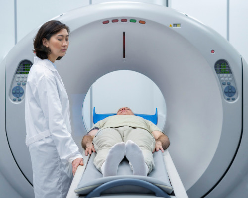
What is an MRI under anesthesia and sedation?
MRI is performed in a strong magnetic field, so preparation begins with a safety check. The doctor asks the patient if they have:
Then, an assessment is made of whether the patient can lie still for the remainder of the examination. If this is difficult, anesthesia for MRI or sedation is discussed—a method that helps perform the examination without unnecessary risk and loss of image quality.
MRI under sedation is performed in a state of controlled sleep or deep relaxation. The patient is not affected by the machine's noise and does not interfere with the scanning process with movement. The depth of exposure is determined after an examination and risk assessment.