MRI of the heart

specialists

equipment

treatment


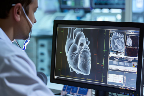
The principle of the tomograph is based on the change in the responses of hydrogen atoms under the influence of a magnetic field. The device reads them and sends them to the computer monitor in the form of achromatic images. Using computer modeling programs, it is possible to create a 3D model of the heart in real time to identify pathological changes and their localization.
The patient is prescribed MRI of the coronary arteries and heart in the following cases:
- Suspected damage to the coronary vessels and aorta, atherosclerosis, rheumatism, endocarditis
- Ischemia
- Inflammatory processes
- Congenital heart defects
- Diagnosis of the myocardium and cardiac neurosis
- Neoplasms in the heart area
- Cardiomyopathy
- Diseases of the aorta
- The need to assess the viability of the myocardium
- Aneurysm and pseudoaneurysm of the ventricles
To obtain more accurate data, MRI with contrast is used. The technique is based on the use of a contrast agent with gadolinium. It is injected into the patient, and during scanning, pathological areas on the image are colored. This allows you to identify the smallest violations, including small tumors, up to several millimeters in diameter.

MRI with contrast is not performed in cases of renal and hepatic insufficiency, as well as in cases of allergic reactions to the drugs used.
Contraindications to MRI diagnostics will be:
- Patient body weight over 120 kg
- Presence of a pacemaker, insulin pump, intracardiac catheter, stent or vascular clip
- Patient has metal implants or tattoos with metal dyes
- 1st trimester of pregnancy
Patients with acute claustrophobia are recommended to take sedative medications before the procedure.
General information
How to prepare for the examination
MRI of the heart vessels does not require special preparation. If the patient is allergic or a similar examination has previously provoked side effects, the attending physician and diagnostician should be notified of this.
Immediately before the procedure, it is necessary to get rid of metal objects (jewelry, accessories) and gadgets.
If possible, you should collect the results of previous examinations and take them with you before going to the cardiology center, where MRI is performed by appointment.


Our doctors


This award is given to clinics with the highest ratings according to user ratings, a large number of requests from this site, and in the absence of critical violations.

This award is given to clinics with the highest ratings according to user ratings. It means that the place is known, loved, and definitely worth visiting.

The ProDoctors portal collected 500 thousand reviews, compiled a rating of doctors based on them and awarded the best. We are proud that our doctors are among those awarded.
Make an appointment at a convenient time on the nearest date
Price

Appointment to the doctor
Reviews


Our clinics

Application “Personal Account K+31”


MRI of the heart and coronary vessels: what the study shows
According to WHO statistics, up to 10% of all heart diseases occur in people under 40 years of age. With age, the number of diseases associated with disorders in the cardiovascular system increases. The situation is worsened by the fact that in the early stages of heart and vascular pathology they do not manifest themselves in any way or are accompanied by non-specific symptoms. This is why accurate diagnostics are so important for the treatment of these diseases.
MRI of the heart and coronary vessels allows you to obtain detailed, layered images of the area being examined. Thanks to this, the doctor can:
Conducting MRI helps in preoperative preparation, treatment planning for hypertrophic cardiomyopathy, and detection of neoplasms.