MR-enterography is now in K+31
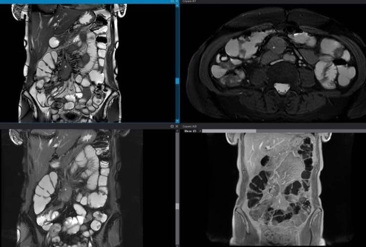
Magnetic resonance imaging (MR enterography) is an accurate and highly informative study that allows you to obtain a detailed image of the small intestine.
MR enterography is an effective way to diagnose diseases of the small intestine, along with endoscopy and computed tomography. Using this diagnostic method, you can accurately determine the state of the mucous membrane of any part of the small intestine and identify pathology at an early stage.
Be sure to read the preparation instructions before examining.
Most often it is carried out in the case of:
- inflammatory diseases of the small intestine;
- masses of the small intestine;
- Crohn's disease;
- obstruction and narrowing of the intestine;
- bleeding from the small intestine;
- abscess of the wall of the small intestine and/or surrounding tissues;
- fistula between bowel loops.
After undergoing MR enterography, the gastroenterologist will give a recommendation for further treatment of the pathology.
The procedure is not recommended if:
- the presence of a pacemaker;
- availability of metal structures;
- pregnancy.
MRI at K+31 has a diagnostic power of 3 Tesla. That is, almost the maximum value available today in diagnostic practice.
Scans provide 360-degree coverage - any type of scan for patients of all sizes.


