MRI of the sacral spine

specialists

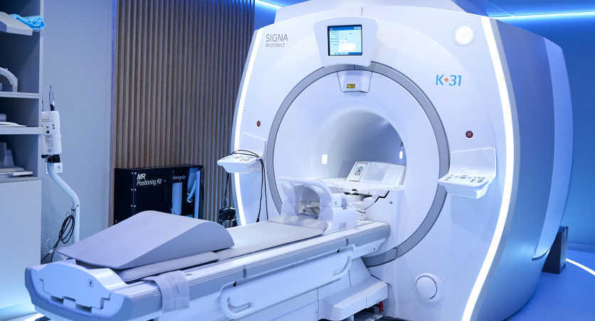
equipment

treatment


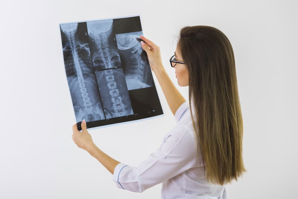
MRI of the sacrum and sacral joints is recognized as the main diagnostic method in orthopedics and rheumatology. The study is carried out by referral from specialist doctors after the initial examination of the patient and the implementation of basic imaging methods (ultrasound, radiography).
Indications for magnetic resonance imaging are the following symptoms:
- Back pain of varying nature and intensity, which can radiate to the intimate area, buttocks, thigh muscles
- History of osteochondrosis, intervertebral hernia
- Sacral injuries
- Suspected degenerative diseases of the nervous system
- Deterioration of blood flow in the vessels of the legs
- Impaired sensitivity of the lower extremities and pelvic area
- Loss of voluntary control over the work of the bladder and intestines
- Erectile dysfunction in men, if typical causes of pathology have already been excluded
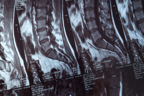
Clear and detailed visualization of the sacral spinal canal during MRI helps the doctor to assess the anatomical features of the area being examined. Such diagnostics are prescribed as part of preparation for surgery in order to plan the tactics of surgical intervention. Tomography is also used for postoperative monitoring of the effectiveness of treatment.
Standard MRI of the sacral spine has a small number of limitations. The main ones are electronic and ferromagnetic implants in the patient's body, which can cause complications and pain during the procedure. Contraindications to magnetic resonance imaging include:
- Implanted pacemaker, cardioverter-defibrillator, prosthetic heart valves
- Fixing structures in bones and joints
- Metal fragments after injuries
- Cochlear implants
- Sewn-in nerve stimulators
- Metal vascular clips on cerebral arteries
MRI of the lumbosacral spine is usually not performed on pregnant women, especially in the first trimester. This is a relative contraindication, therefore, if there is an urgent need, diagnostics are performed taking into account possible risks.
Tomography with contrast is not prescribed to patients with gadolinium intolerance, polyvalent drug allergy, renal failure and decompensated bronchial asthma.
General information
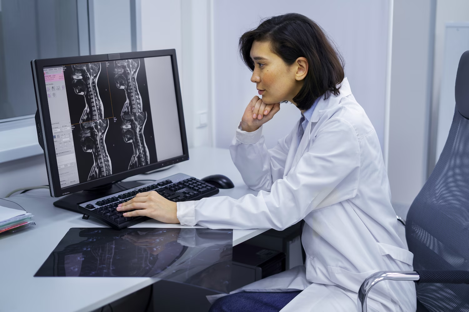
How is the procedure performed?
The examination does not require any special preparation. If a contrast agent is planned to be administered, you should refrain from eating for 2-3 hours. Before the examination, you will need to remove all jewelry and clothing with metal parts.
The scanning is performed with the patient lying on his back. The limbs are fixed to exclude spontaneous movements, and the head will need to be placed on a special stand. Then the table moves into the tomograph, and the magnetic resonance imaging begins. The diagnosis is not accompanied by pain or other unpleasant sensations, and headphones are provided to protect against the noise of the operating device.
MRI of the lumbosacral spine is quick - about 20 minutes without contrast and 15 minutes longer if contrast is administered during the procedure. The patient can leave the clinic immediately after the end of the examination.
Our doctors


This award is given to clinics with the highest ratings according to user ratings, a large number of requests from this site, and in the absence of critical violations.

This award is given to clinics with the highest ratings according to user ratings. It means that the place is known, loved, and definitely worth visiting.

The ProDoctors portal collected 500 thousand reviews, compiled a rating of doctors based on them and awarded the best. We are proud that our doctors are among those awarded.
Make an appointment at a convenient time on the nearest date
Price
Other services

Appointment to the doctor
Reviews


Our clinics

Application “Personal Account K+31”


Diagnostic value of MRI
The technology works on the principle of irradiation with electromagnetic waves that cause resonance in tissues, after which the signals are captured and analyzed by a computer system. The study provides clear images of soft tissues, cartilage, blood vessels and nerves, unlike CT, which more clearly visualizes bone structures.
MRI of the sacral spine shows:
Additionally, patients may be prescribed MRI of the sacroiliac joints (SIJ) - this is the only reliable method for confirming the diagnosis sacroiliitis. The technique shows inflammatory changes, pathological fatty infiltration of the bone marrow, sclerotic processes and ankylosis of the joints.