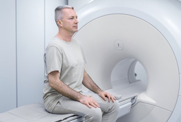
MRI of the lumbar spine
Magnetic resonance imaging is considered the safest method for diagnosing pathologies of internal organs, due to the absence of radiation or radio wave exposure and a small number of contraindications. The study of the lumbosacral spine (MRI LS) is carried out to identify structural and functional disorders in the vertebrae, joints, sacrum, surrounding soft tissues, nerves and vessels of this area of the back.

specialists

equipment

treatment


When is MRI of the POP prescribed?

Magnetic resonance imaging of the lumbosacral region is prescribed for:
- Frequent pain in the lumbar region, radiating to the leg or pelvic region
- Weakening or loss of sensation and reflexes in the legs
- Spinal injuries
- Pelvic organ dysfunction
- Chronic or acute inflammation of the lumbar tissues
- Suspected oncology of bone tissues of the lumbar region
- Long-term muscle weakness in the legs
- Syringomyelia or multiple sclerosis
- Suspicion of intervertebral hernias
- Symptoms of osteochondrosis
- Anomalies of the spine structure
- Disturbance of blood circulation in the lower extremities
Contrast-enhanced MRI is used to detect tumors or metastases. After the introduction of a special substance, blood vessels are visualized in contrast to stationary tissues, which allows for clearer images of veins, arteries, and capillaries.
General information
What does an MRI of the lumbar spine show?
Lower back pain can be associated with spinal or pelvic trauma, or be a consequence of diseases of the back or internal organs.
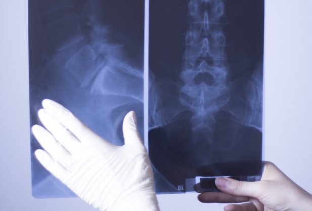
A regular X-ray is not always enough for a doctor to make a diagnosis.
If a specialist has given a referral for MRI of the POP, what can it show? Diagnostics are carried out for the purpose of:
- Finding out the cause of back pain to determine treatment tactics
- Detection of neoplasms in the sacrum and adjacent tissues, assessment of their nature (benign or malignant)
- Determining the presence of distant tumor metastases in the lumbar region
- Detection of intervertebral disc herniations and their sizes
- Research into the area of nerve root damage
MRI provides a 2-3 dimensional graphic image of the spine. Thanks to this, the doctor receives accurate information about:
- Structural disorders in the vertebrae, intervertebral joints
- The presence and characteristics of protrusions, hernias, injuries to bone structures, ligaments and cartilage
- Degrees of soft tissue damage
- Locations of pinched nerves
- Functional changes in blood vessels, spinal cord
With the help of tomography, it is possible to make an accurate initial diagnosis, as well as track the dynamics of the patient's condition after treatment.

Our doctors


This award is given to clinics with the highest ratings according to user ratings, a large number of requests from this site, and in the absence of critical violations.

This award is given to clinics with the highest ratings according to user ratings. It means that the place is known, loved, and definitely worth visiting.

The ProDoctors portal collected 500 thousand reviews, compiled a rating of doctors based on them and awarded the best. We are proud that our doctors are among those awarded.
Make an appointment at a convenient time on the nearest date
Price

Appointment to the doctor
Reviews


Our clinics

Application “Personal Account K+31”


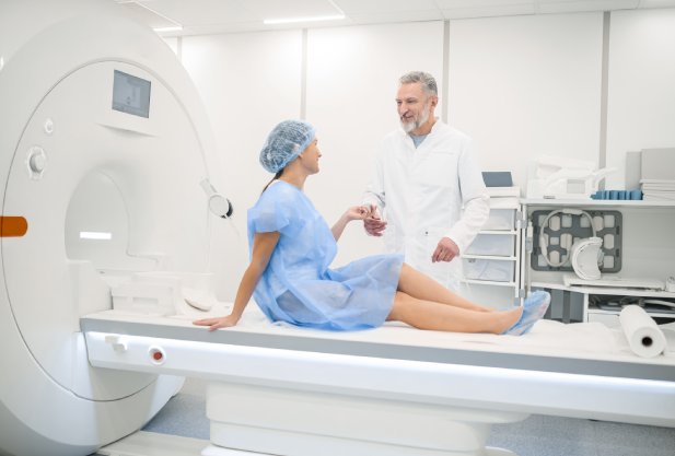
Types of MRI of the lumbar spine
Tomography of the lumbar region can be performed in two ways:
The method of conducting diagnostics is determined by the attending physician - therapist, traumatologist, neurologist, surgeon or other specialist who issued the referral for the examination. The choice of method depends on the preliminary diagnosis, individual indications and contraindications, anamnesis and general condition of the patient.