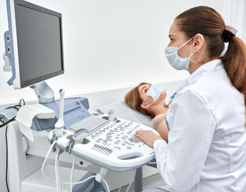
Ultrasound (ultrasound diagnostics)
The K+31 ultrasound diagnostics department operates across three clinics in Moscow, bringing together specialists with expertise in cardiology, hepatology, obstetrics, oncology, and vascular pathology. There is no such thing as a "routine ultrasound" here; every examination is performed by a physician specializing in the specific area relevant to your condition—a doctor ready to answer clinical questions on the spot.
Urology
- Ultrasound of the kidneys, bladder, and ureters
- Transrectal ultrasound (TRUS) of the prostate with ultrasound-guided biopsy capability
- Scrotal ultrasound with Doppler imaging
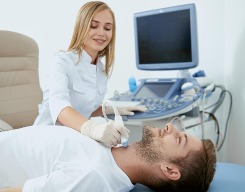
Cardiovascular System
- Echocardiography (EchoCG): standard, stress EchoCG, transesophageal
- Duplex scanning of carotid and vertebral arteries, and veins and arteries of the upper and lower limbs
- Transcranial Doppler (TCD)
- Penile artery Doppler ultrasound
- Ultrasound of abdominal vessels (portal vein, renal arteries, abdominal aorta)
Endocrinology
- Ultrasound of the thyroid and parathyroid glands
- Adrenal gland ultrasound
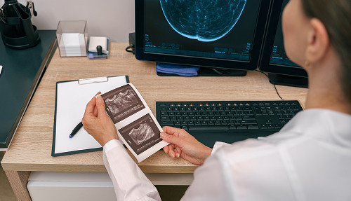
Mammology
- Breast ultrasound with Doppler imaging
- Joint protocol with a mammologist: immediate consultation with a K+31 breast oncologist upon detection of abnormalities
Musculoskeletal System
- Ultrasound of joints, soft tissues, tendons, and ligaments
- Ultrasound guidance for intra-articular injections
Specialized Examinations
- Shear-wave elastography (SWE) of the liver — non-invasive assessment of fibrosis using the METAVIR scale
- Ultrasound-guided biopsy (liver, thyroid gland, mammary gland, prostate)
- Ultrasound of the pleural cavities
- Ultrasound of lymph nodes
- Neonatal neurosonography
- Home ultrasound — mobile team visit
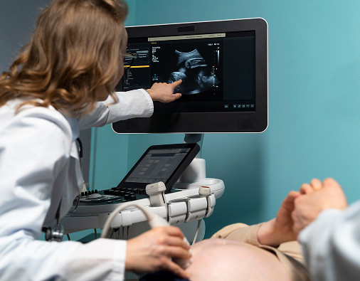
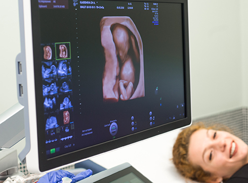
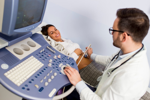
According to the International Society of Ultrasound in Obstetrics and Gynecology (ISUOG), the best ultrasound diagnostic system for performing ultrasound during pregnancy in all trimesters is the General Electric Voluson E10, which our clinic also has.
We offer expectant mothers ultrasound examination of the fetus at various stages of pregnancy. Using our equipment, it is possible to conduct screening to assess intrauterine development.
Doppler ultrasound of the uteroplacental and fetal blood flow has important diagnostic value in the group of pregnant women at high perinatal risk.
Ultrasound is quite informative for multiple pregnancies, various pathologies, and late pregnancy. The research method allows us to identify pathological changes and anomalies in fetal development.
Timely detection of violations makes it possible to prevent the development of severe complications of pregnancy and develop tactics for managing a pregnant woman.
Indications for ultrasound
An ultrasound is prescribed when it's necessary to accurately determine the cause of health problems. For example, if you have abdominal pain, your doctor will recommend an examination to rule out serious conditions like kidney stones or inflammation.
An ultrasound is also useful, for example, if a tumor is suspected—an examination can be performed to rule out or confirm the presence of a growth, determine its size, and determine its location.
Another important indication for an ultrasound is pregnancy. Expectant mothers regularly undergo ultrasounds to ensure the baby is developing normally and there are no threats.
This test is also often used for heart and vascular problems, as it allows one to detect blood clots, thickening of the walls, or circulatory problems.
Endocrine system complaints are also a reason to see a specialist. If thyroid dysfunction is suspected, it's best to immediately undergo an ultrasound to initiate treatment promptly. Therefore, the primary indication for an ultrasound is the need for an accurate diagnosis of the patient's health.
PREPARATION FOR ULTRASOUND
Ultrasound of the abdominal organs, renal vessels, and abdominal vessels
(abdominal aorta and its visceral branches, portal vein and its branches, inferior vena cava and its branches, iliac arteries and veins).
Preparation: 24 hours before the examination, exclude fresh fruits and vegetables, carbonated drinks and juices, legumes, baked goods, milk, and brown bread from your diet. Arrive for the examination strictly on an empty stomach (do not eat or drink). It is advisable to refrain from smoking for 2 hours before the examination. Diagnostic procedures such as gastroscopy, colonoscopy, and barium studies of the gastrointestinal tract should not be performed before an abdominal ultrasound (they are performed only after the ultrasound).
Abdominal ultrasound with a choleretic breakfast
Preparation is similar to that for a standard abdominal ultrasound, plus you must bring a choleretic breakfast, which you will need to eat after the initial examination.
Choleretic breakfast (options):
- 20% sour cream: 200–250 g (at least 20% fat content)
- 10% yogurt: 200–250 ml (at least 10% fat content)
- 20% cream: 200–250 ml (at least 20% fat content)
Ultrasound of the bladder, prostate gland (transabdominal), pelvic organs in women (transabdominal)
The ultrasound is performed with a full bladder — the urge to urinate should be felt at the time of the examination.
To quickly fill your bladder: first drink coffee or green tea, then 3-4 glasses of water – your bladder will usually fill in 20-30 minutes.
Pelvic Ultrasound in Women (Transvaginal Access)
The ultrasound is performed with an empty bladder – you must urinate before the examination.
Ultrasound of the Lower Extremity Vessels (Arteries or Veins)
The examination is performed starting from the groin area. On the day of the examination, wear comfortable, stretchy underwear.
The clinic uses the Philips Affinity 70 expert-class ultrasound machine to perform liver shear wave elastometry. Diffuse liver diseases are one of the biggest problems worldwide. The etiology of these diseases is varied:
- Viral hepatitis (hepatitis B or C).
- Fatty liver infiltration of alcoholic and non-alcoholic origin.
- Autoimmune hepatitis.
- Medication-related complications.
- Primary biliary cirrhosis and other less common pathological conditions and diseases.
Our clinic uses recommendations developed by the Russian Association of Ultrasound Specialists in Medicine and the Department of Ultrasound Diagnostics at the University of Pavia (Italy) for assessing liver stiffness, primarily in the setting of viral hepatitis C. Shear wave elastography is based on measuring the propagation velocity of a shear wave and allows the specialist to obtain a digital liver stiffness value, which can be correlated with various assessment scales, including the METAVIR scale.
General information
How the examination is conducted
- Booking and consultation — when booking online or by phone, the receptionist clarifies the purpose of the examination and, if necessary, refers you to the appropriate ultrasound specialist
- Preparation — specific to each type of examination (see the "Preparation" section)
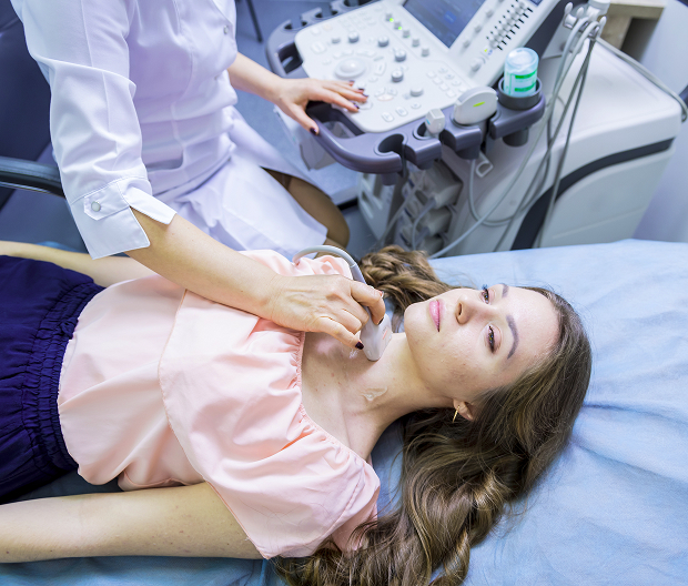
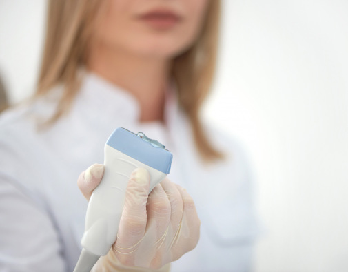
- Procedure — the patient lies comfortably on the exam table; the doctor applies acoustic gel and moves the transducer; the process takes 15–40 minutes depending on the scope of the examination
- Report — issued immediately upon completion or within 30 minutes for complex cases; available in your personal account on the website
- Results consultation — the doctor explains any findings and, if necessary, arranges a referral to a relevant specialist

Answers to frequently asked questions
What ultrasound technologies do K+31 doctors use?
K+31 Clinic is equipped with expert-class stationary and portable ultrasound systems from leading manufacturers (Toshiba, General Electric, Philips, Hitachi), offering:
- A wide range of diagnostic capabilities
- Excellent visualization of organ and tissue structures
- Doppler imaging programs
- Panoramic scanning
- Real-time 3D imaging
The premium-class ultrasound systems installed at the clinic—including the Canon i800, Toshiba Artida, and Toshiba Aplio 500—incorporate innovative imaging technologies and unique capabilities. These systems deliver unprecedented grayscale image quality with exceptional detail regarding tissue and organ structures.
Advanced computer processing capabilities for 3D/4D imaging on these latest-generation systems allow for the creation of spatial reconstructions of organs, blood vessels, and pathological lesions.
Our state-of-the-art equipment enables the diagnosis of pathologies affecting the urinary and reproductive systems.
Which organs and systems can be examined via ultrasound at K+31?
K+31 performs virtually all currently available ultrasound examinations:
- Brain
- Abdominal organs
- Kidneys and adrenal region
- Liver
- Thyroid gland
- Breasts
- Salivary glands
- Lymph nodes
- Lungs
- Pleural cavities
- Eye orbits
- Pelvic organs (male and female)
- Urinary bladder
- Scrotal organs
- Prostate gland
- Soft tissues and joints
- Abdominal cavity
- Gallbladder
- Retroperitoneal space
- Pancreas
- Spleen
- Lower extremities
- Upper extremities
- Heart (Echocardiography)
- Blood vessels
- Triplex and duplex scanning of major vessels, veins, and visceral arteries
- Brachiocephalic artery scanning (with color Doppler flow mapping and rotational maneuvers)
- Doppler echocardiography, stress echocardiography, and transesophageal echocardiography
- Transcranial Doppler (TCD)
- Ultrasound diagnostics during pregnancy (all trimesters)
- Transvaginal examination of the uterus and cervix (first trimester)
- Doppler assessment of uteroplacental and fetal blood flow
- 3D/volumetric imaging
- fetus
- Neurosonography from the first day of life
- Ultrasound guidance for minimally invasive interventions
What types of ultrasound are there?
Depending on the medical condition, the attending physician may prescribe the following types of ultrasound examinations:
- Densitometry is a diagnostic method involving the scanning of bones using ultrasound or X-rays. Recently, this examination has become particularly relevant for patients who have recovered from COVID-19. Densitometry is often prescribed in conjunction with other tests.
- Doppler ultrasound (Dopplerography) is an examination used to assess blood flow. The specialist evaluates the veins and arteries of the upper and lower limbs, the brain, and the pelvic organs. It is also prescribed during pregnancy to detect cardiovascular abnormalities in the fetus.
- Sonography is a less common term for Doppler ultrasound.
- 3D ultrasound is a diagnostic procedure that provides a more precise image of internal tissues and organs. This medical examination allows for a three-dimensional view, facilitating disease diagnosis and treatment planning. 3D and 4D ultrasounds are frequently prescribed during pregnancy.
- Duplex scanning is an ultrasound procedure used to examine the blood vessels of the neck and brain, as well as the veins of the lower limbs. The method combines Doppler ultrasound with B-mode imaging, allowing for the visualization of veins, arteries, and surrounding soft tissues.
How do you interpret an ultrasound report?
How does an ultrasound at K+31 differ from one at a standard local outpatient clinic?
Three key differences. First, the equipment: expert-class systems from GE, Philips, and Canon, as opposed to standard mid-range units. Second, physician qualifications: at K+31, ultrasound specialists hold specific medical certifications and conduct examinations exclusively within their area of expertise. Third, clinical integration: any detected pathology is immediately incorporated into the patient’s care pathway within the clinic, eliminating the need to book a new appointment or explain the situation from scratch.
Do I need a doctor's referral to book an ultrasound?
No. You can book an appointment yourself, without a referral. If you are unsure which type of examination you need, a K+31 receptionist or the on-duty consulting physician will help you decide.
How soon will the results be ready?
The examination report is issued immediately after the procedure—typically within 15–30 minutes. In complex cases (for example, if an atypical growth is detected), the doctor may consult a colleague; the result will be ready later that same day. The report is available in your personal account on the K+31 website or in the app.
Is it possible to have several types of ultrasound scans during a single visit?
Yes. When booking your appointment, please inform the receptionist that you need multiple examinations; the specialist will coordinate the timing and any necessary preparation. Certain combinations (for example, abdominal, kidney, and thyroid ultrasounds) can be performed in a single session.
Do children undergo ultrasound examinations?
Yes. Neonatal neurosonography, abdominal and pelvic ultrasound for children of all ages, and echocardiography are all available at K+31. For gynecological examinations of girls and adolescents, only the transabdominal method is used.
Is the branch open on weekends?
Yes. K+31 clinics are open on Saturdays and Sundays from 9:00 AM to 6:00 PM or 7:00 PM (depending on the branch). Certain emergency tests are available around the clock. Please check the schedule for the specific branch when booking an appointment.
What is liver elastography, and how does it differ from a standard ultrasound?
Standard liver ultrasound assesses the organ's structure and size and detects focal lesions. Elastography additionally measures tissue stiffness—a mechanical parameter reflecting the degree of fibrosis. This enables non-invasive staging of fibrosis (without a biopsy) according to the METAVIR scale: from F0 (normal) to F4 (cirrhosis). It is particularly valuable for monitoring chronic hepatitis and tracking treatment progress.
Can I get ultrasound results online?
Yes. The report and images are saved to your electronic medical record at K+31 and are accessible via your personal account on the k31.ru website. If necessary, the results can be sent via email or a messaging app.
Reviews
Our doctors


This award is given to clinics with the highest ratings according to user ratings, a large number of requests from this site, and in the absence of critical violations.

This award is given to clinics with the highest ratings according to user ratings. It means that the place is known, loved, and definitely worth visiting.

The ProDoctors portal collected 500 thousand reviews, compiled a rating of doctors based on them and awarded the best. We are proud that our doctors are among those awarded.
Make an appointment at a convenient time on the nearest date
Price

To get a consultation


Our clinics

Application “Personal Account K+31”


What is an ultrasound?
Wide Range of Examinations
K+31 offers a full range of modern ultrasound diagnostic procedures—from routine abdominal scans to complex ultrasound-guided interventional procedures:
Abdominal and Retroperitoneal Organs
Gynecology and Obstetrics