Renal colic: how is it treated in a hospital setting?
Renal colic usually begins suddenly. It is a sharp pain in the side or lower back that makes it difficult for a person to function normally. Nausea, sweating, and a fever occur.
The main cause of the pain is urolithiasis (urolithiasis): a calculus (stone) becomes lodged in the ureter and prevents urine from passing naturally. This causes the kidney to become distended.
Renal colic is treated in a hospital setting. Hospitalization is necessary not only for pain relief but also to rule out dangerous complications such as obstruction, hydronephrosis, and infection.
According to ICD-10, renal colic is classified under code N23. Inpatient care is based on clinical guidelines: pain relief begins, then the stone's location is determined and a treatment method is chosen.

specialists

equipment

treatment


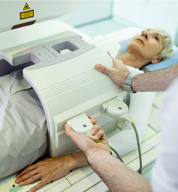
Diagnostics in the emergency room and hospital
The hospital physician's job is to confirm that the patient has renal colic. If inflammation is present, a decision is made about treatment: conservative treatment (medication) or surgery.
Laboratory tests (urine and blood tests)
The following tests are taken in the hospital for renal colic:
- Urinalysis
- Blood analysis
- Blood chemistry (creatinine, electrolytes)
Based on the results, the degree of inflammation is assessed and a decision is made about what to do next: surgery or waiting.
Instrumental methods (ultrasound, CT, excretory urography)
The gold standard for diagnostics today is CT. Only a tomography scan can accurately determine the stone's location, size, and density.
Ultrasound is rarely used: it's virtually impossible to visualize the stone.
Excretory urography is also performed. However, this test is reserved for patients with suspected ureteral obstruction.
Inpatient treatment for renal colic begins with an examination. It's impossible to visually determine the presence of a stone or its size.
Stages of inpatient treatment
First, they relieve the pain and spasm, then decide how to pass the stone without damaging the kidney. Kidney stone treatment is a lengthy process, so don't expect a quick recovery.
Relief of an acute pain attack
First, relieve the renal colic, then everything else. Antispasmodics and nonsteroidal drugs are used to relieve pain. Pulse and blood pressure are monitored during treatment.
Important: renal colic cannot be completely relieved. It is only necessary to reduce the severity of the pain; otherwise, important symptoms may be missed.
Conservative lithokinetic therapy (stone expulsion)
If the stone is small, there is no infection, and the kidney is not critically damaged, a watchful waiting approach with lithokinetic therapy may be used. At this stage, doctors control pain and monitor urine output.
For larger kidney stones, a different treatment is prescribed. Doctors first try to dissolve the stone with medications. If successful, the stone will pass on its own in the urine.
Surgical methods for removing stones
If the pain persists, kidney stones need to be removed. This procedure is also performed if the stone is lodged in the ureter.
To allow urine to drain naturally, a stent or drainage is placed. All these procedures are performed only in a hospital setting.
Modern minimally invasive surgeries
Today, most interventions for urolithiasis are performed without open surgery. The choice of method depends on the size, density, and location of the stone (ureter, renal pelvis), as well as the patient's age and health.
Extracorporeal shock wave lithotripsy (ECS)
Extracorporeal shock wave lithotripsy is the fragmentation of stones using a shock wave without incisions. This procedure is not prescribed to all patients: it is only recommended for those with soft stones (the density is determined by CT).
The stone fragments are not removed from the ureter; they are passed naturally.
Contact laser lithotripsy
Laser stone fragmentation is performed through a thin endoscope inserted into the ureter. The stone is crushed by the laser, and the fragments are removed. This is also a relatively simple procedure. It is suitable for crushing denser stones. Anesthesia is administered for this procedure.
Percutaneous nephrolithotripsy
Nephrolithotripsy is used for large kidney stones, especially if they occupy the renal pelvis or several calyces. In this case, a puncture is essential. The surgeon enters the renal pelvis through the lower back, crushes the stone, and completely removes its components. This is a more serious procedure, requiring a longer hospital stay.
Table: Stone crushing methods
| Method | Invasiveness | Effectiveness for hard stones | Recovery time |
|---|---|---|---|
| Extracorporeal radiation lithotripsy (ESRL) | Non-invasive | Moderate | Often outpatient |
| Contact laser lithotripsy | Minimally invasive | High | 1–2 days in hospital |
Indications for discharge and rehabilitation

The patient is discharged after full recovery. Although most stones are removed without punctures, the patient still spends 5 to 10 days in the hospital. Many patients are given IVs during this time.
Upon discharge, the doctor explains the fluid regimen and how to control urination.
FAQ
Is it possible to wait out the colic at home if the pain has subsided?
Is kidney stone removal always necessary?
Why do they perform stenting if the stone still needs to be crushed?
How is renal colic treated?
Caution: If you suspect inflammation, consult a doctor or call an ambulance. Do not apply heat to the painful area or take a bath.
Our doctors


This award is given to clinics with the highest ratings according to user ratings, a large number of requests from this site, and in the absence of critical violations.

This award is given to clinics with the highest ratings according to user ratings. It means that the place is known, loved, and definitely worth visiting.

The ProDoctors portal collected 500 thousand reviews, compiled a rating of doctors based on them and awarded the best. We are proud that our doctors are among those awarded.
Make an appointment at a convenient time on the nearest date
Other services

Appointment to the doctor
Reviews


Our clinics

Application “Personal Account K+31”


Why is hospitalization necessary for renal colic?
Renal colic causes severe pain. But this isn't the only reason to see a doctor. The pain may be due to urinary tract obstruction. If urine doesn't flow, pressure in the renal pelvis increases, and hydronephrosis can develop. As a result, kidney function deteriorates, and intoxication occurs.
A second complication is infection. In this case, the condition can quickly become severe.
There are situations when emergency hospitalization for renal colic is necessary. Call an ambulance if you experience the following symptoms:
Pregnant women, people with a single kidney, and transplant patients should call an ambulance at the slightest sign of colic.
Avoid warming the lower back or taking a hot bath: heat accelerates the spread of infection and worsens the condition.