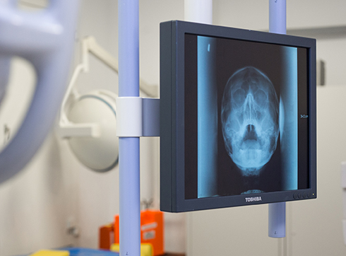
Doppler ultrasound of the head and neck vessels
One of the most effective non-invasive diagnostic methods is Doppler ultrasound. The examination requires no special preparation and is suitable for virtually everyone, regardless of age or health. Doppler ultrasound allows, among other things, for the analysis of the vessels of the neck and head.

specialists

equipment

treatment


Indications for Ultrasound Doppler Imaging of the Head and Neck Vessels
A general practitioner, neurologist, neurosurgeon, or other specialist doctor can refer a patient for an ultrasound examination of the head and neck vessels. This method allows for the detection of a wide range of vascular problems, down to the smallest disturbances in blood flow. Simply put, the procedure gives doctors a clear picture of the condition of the blood vessels.
This test is often prescribed for conditions that increase the risk of vascular complications. Here are some of them:
- Cervical osteochondrosis
- Disorders of the nervous system that regulates blood circulation
- Vascular atherosclerosis
- History of strokes or transient ischemic attacks
- History of traumatic brain injury
- Blood clots in blood vessels
- Blood clotting problems
A referral for such diagnostics may also be made before heart surgery or major interventions to assess risks in advance.
Furthermore, complaints such as the following may be grounds for investigation:
- Sleepless nights or frequent awakenings in the dark
- Regular headaches of varying intensity
- Constant dizziness
- Migraine attacks
- Tingling or weakness in the arms and legs
- Rapid fatigue even with minor exertion
- Tinnitus
- Hearing loss
- Decreased visual acuity, appearance of "spots" before the eyes
- Fainting or pre-fainting spells
- Sudden changes in well-being depending on the weather
- Memory impairment and decreased ability to concentrate
Vascular examinations can be performed not only for specific diseases but also in the presence of symptoms indicating insufficient blood supply to the brain.
Contraindications to Doppler ultrasound of the vessels of the head and neck
Doppler ultrasound of the vessels of the head and neck is a highly safe procedure and is suitable for virtually every patient. This technique is so versatile that there are simply no absolute contraindications.
However, there is one time limitation related solely to the condition of the skin in the area being examined. If there are fresh scratches, open wounds, irritation, or inflammation of the skin in the neck or temple area, the Doppler ultrasound is temporarily postponed. The device's sensor must fit snugly against the skin to ensure good contact, and tissue damage interferes with the normal transmission of ultrasound waves.
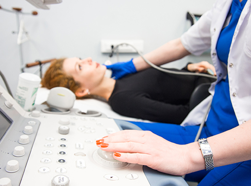
How is the examination of the vessels of the head and neck performed?

The examination takes place in a specialized medical room equipped with the necessary equipment and sensors. The doctor asks the patient to assume a comfortable position on a medical bed, most often horizontal. For comfort, a small pillow is used under the head, and for those with a short neck, a soft cushion is placed between the shoulder blades. This position ensures maximum accessibility for the diagnostician to the areas being examined.
Before the procedure, the patient must inform the specialist of any existing health problems that could affect the transmission of ultrasound signals. This typically includes diagnoses such as cervical osteochondrosis, intervertebral disc disease, ankylosing spondylitis, unstable vertebrae, and other musculoskeletal conditions that can compress blood vessels and distort the true picture.

The examination itself is performed by applying a special gel to the skin, where a sensitive probe is then placed. The gel improves the transmission of ultrasound vibrations, facilitating the acquisition of a clear signal. The physician then carefully moves the probe over the areas of interest, carefully observing the image on the monitor screen. It is important to remain completely calm and avoid talking, as any movement can interfere with the accuracy of the measurement.
An image of the vessel's condition is displayed directly during the examination, allowing the specialist to instantly assess the situation and record the necessary values. The average duration of the entire procedure is approximately 15 minutes. A short additional time is required to prepare a detailed report on the examination results. At the patient's request, a recording of the diagnostic process can be saved to a digital device, facilitating further discussion of the diagnosis with the consulting physician or the use of the obtained data in the comprehensive treatment of complex medical cases.
General information
- Preparation for ultrasound dopplerography of the head and neck vessels
- Results of ultrasound dopplerography of the vessels of the head and neck
- Types of ultrasound dopplerography of the vessels of the head and neck
- Advantages of ultrasound dopplerography in clinics
- Prices for ultrasound dopplerography of the head and neck vessels in Moscow
Preparation for ultrasound dopplerography of the head and neck vessels
The Doppler ultrasound procedure does not require any special preparation, but there are some recommendations to help improve the accuracy of the results. For example, it is advisable to limit cigarette smoking and consuming caffeinated beverages, such as tea, coffee, and energy drinks, 24 hours before the appointment. These measures eliminate the potential impact of temporary factors on the blood vessels, allowing the doctor to form an accurate picture of the patient's health.


Our doctors


This award is given to clinics with the highest ratings according to user ratings, a large number of requests from this site, and in the absence of critical violations.

This award is given to clinics with the highest ratings according to user ratings. It means that the place is known, loved, and definitely worth visiting.

The ProDoctors portal collected 500 thousand reviews, compiled a rating of doctors based on them and awarded the best. We are proud that our doctors are among those awarded.
Make an appointment at a convenient time on the nearest date
Price
Other services

Appointment to the doctor
Reviews


Our clinics

Application “Personal Account K+31”


What is ultrasound dopplerography and its purpose?
UZDG stands for vascular Doppler ultrasound, also known as Doppler analysis. Simply put, it's a test that helps visualize how blood flows through the body's vessels. Doctors use a special device, similar to a scanner, to examine the condition of arteries and veins, identifying possible blood flow problems or vascular narrowing.
The goal of the procedure is simple: to detect circulatory problems early to prevent serious diseases of the heart, brain, and other organs. This test is painless, quick, and completely safe.