Duplex scanning of the brachiocephalic arteries (DS BCA)

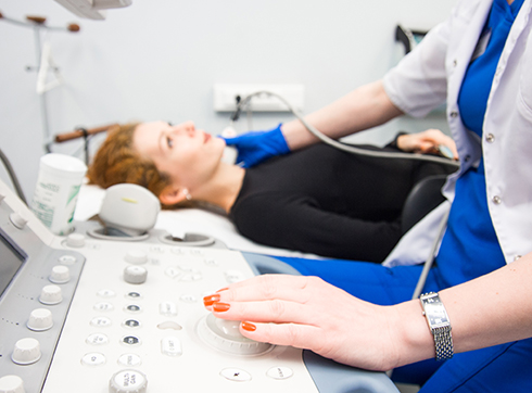
Duplex scanning is a modern ultrasound technique that allows for a highly accurate assessment of the condition of the major vessels supplying the brain. This examination combines visualization of the anatomical structure of the arteries and simultaneous analysis of blood flow characteristics in real time. This combination provides the specialist with comprehensive information about the presence of even the earliest pathological changes, which is important for the early prevention of acute cerebrovascular accidents. The procedure is considered completely safe and does not involve radiation exposure, making it suitable for a wide range of patient groups.

At the K+31 Clinic in Moscow, the examination is performed using expert-class equipment, guaranteeing the highest level of detail and accuracy. Diagnostics are performed by highly qualified physicians with extensive experience in vascular ultrasound. We combine cutting-edge medical technology with affordable prices, providing our patients with confidence in their health and high-quality diagnostics without overpaying.

Best
specialists
Expert
equipment
Advanced diagnostic
treatment
Sign up
Fill out the entry form on the page and we will call you back
Sign up Позвонить
Download the application “Personal Account K+31”
Everything about your health in one click!
Subscribe to social networks K+31
To keep up to date with current offers and receive useful advice about your health.
doctor k31

The essence of the technique and its principle of operation

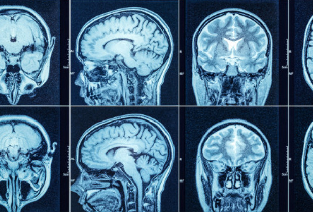
The abbreviation BCA stands for brachiocephalic arteries (BCA), the key main vessels supplying blood to the brain and soft tissues of the head. These include the carotid, vertebral, and subclavian arteries, which form the so-called circle of Willis at the base of the brain—a unique ring-shaped structure responsible for the uniform distribution of blood flow. This technique allows for a detailed assessment of the condition of the extracranial sections of the brachiocephalic arteries located in the neck. The functioning of the central nervous system directly depends on their proper functioning, and any obstruction of these pathways can lead to serious neurological complications, such as ischemic stroke.

Duplex scanning is based on a synthesis of two diagnostic modes. The first, B-mode, or two-dimensional grayscale ultrasound, allows for the visualization of the arteries themselves in real time. The physician sees not just an outline on the monitor, but the detailed structure of the vascular wall: its thickness, integrity, the presence of atherosclerotic plaques, thrombi, or congenital malformations. Essentially, they obtain an ultrasound map of the vascular anatomy.

The second component of the study, spectral Doppler ultrasound, evaluates the functional characteristics of blood flow in the section being examined. Using this physical phenomenon, the device captures the speed and direction of blood flow through the vessel, converting this data into a graph. This allows for hemodynamic analysis: identifying areas of narrowing (stenosis), where flow velocity changes abnormally, or assessing the degree of resistance to blood flow. The color mapping function clearly visualizes multidirectional blood flows on the screen, simplifying the diagnosis of turbulent flow in areas of constriction.

The main advantage of the scan is the simultaneous display of both modes on a single screen. The specialist not only observes the anatomical features of the artery but also analyzes blood flow within its lumen in real time, accurately determining how the identified structural changes affect the blood supply to the brain. This comprehensive approach ensures an accurate diagnosis and the selection of effective treatment strategies.

Why is the procedure performed and how does it differ from other methods?

Doppler ultrasound examination of the brachiocephalic arteries is prescribed for a wide range of diagnostic purposes. It provides the physician with comprehensive information that allows them not only to detect the presence of a disease but also to determine its stage, potential risks, and progression.

The main objectives of the examination are:

  1. Assessment of the structural condition of the vessels. The physician examines the structure of the vascular wall in detail and measures its thickness, which is a key marker for the onset of atherosclerotic changes before plaque formation.
  2. Detection of atherosclerotic lesions. The examination allows the detection of cholesterol plaques, their size, structure (stable or unstable), and the extent to which they obstruct the artery and impair blood flow.
  3. Diagnosis of deformities and anomalies. This method effectively detects pathological tortuosity, looping, and other congenital or acquired structural anomalies of the brachiocephalic arteries.
  4. Detection of stenoses and occlusions. The procedure accurately determines the presence and degree of narrowing or complete occlusion of the vessel, which is crucial for predicting stroke risk.
  5. Blood flow analysis. Doppler ultrasonography measures the speed and direction of blood flow and evaluates vascular resistance, providing a comprehensive picture of the quality of cerebral blood supply.

As for the advantages of this technique, it is rightfully considered the gold standard in the primary diagnosis of pathologies of the arteries supplying the brain.

Key advantages:

  1. Absolute safety. The method is based on ultrasound, which does not emit ionizing radiation. This makes the examination suitable for repeated use to monitor the condition over time, as well as for a wide range of patients, including the elderly and those with comorbidities.
  2. Non-invasive and painless. The procedure does not require breaking the skin, injections, or insertion of instruments. It is comfortable for the patient and causes no discomfort.
  3. Highly informative. Modern ultrasound scanners provide excellent visualization, allowing for detailed data on both vascular structure and functional indicators of blood circulation.
  4. No preparation required and quick. The examination requires no complicated preparation and takes an average of 20 to 40 minutes.
  5. Speedy results. The ultrasound technician can provide a report immediately after the procedure, allowing the attending physician to quickly make a diagnosis and prescribe treatment.

In what cases is it recommended to undergo an examination?

Duplex scanning is a reasonable procedure in a number of clinical situations and risk factors. This examination provides an objective picture of the condition of the major vessels and assesses the risk of developing cerebrovascular pathologies.

Diagnostics are especially important in the following cases:

  1. Presence of risk factors and chronic diseases. The procedure is indicated for patients with arterial hypertension, diabetes, elevated cholesterol levels, and diagnosed atherosclerosis of other vascular beds. These conditions greatly increase the likelihood of damage to the brachiocephalic arteries.
  2. Planning major surgical interventions. The examination is often prescribed as part of preoperative preparation for cardiac procedures, such as coronary artery bypass grafting. This allows for risk assessment and the prevention of severe brain complications during surgery.
  3. Dynamic monitoring of vascular health. For patients with already diagnosed pathologies, such as stenosis or atherosclerotic plaques, duplex scanning becomes the primary tool for monitoring the progression of the disease and the effectiveness of prescribed therapy.
  4. Post-operative monitoring. The study is indispensable for monitoring the results of surgical treatments on neck vessels, such as stenting and carotid endarterectomy. It allows for the timely detection of possible complications, such as restenosis.

Warning signals requiring specialist attention

The appearance of neurological symptoms indicating a possible disruption of cerebral blood flow should prompt immediate medical attention and subsequent duplex scanning.

Such warning signs include:

  • Episodes of sudden dizziness, especially associated with head movements or changes in body position
  • Spontaneous tinnitus, which may be constant or intermittent
  • Unexplained headaches, which are not clearly localized and often diffuse
  • Visual disturbances: the appearance of a veil, spots, short-term loss of visual field
  • Sudden and unexplained weakness
  • Unsteadiness of gait, loss of coordination
  • History of transient ischemic attacks, which manifest as temporary weakness in the limbs, numbness, and speech impairment

General information

Restrictions on the diagnostic procedure

Unlike many diagnostic methods, duplex arterial scanning is characterized by a complete absence of absolute contraindications. This is directly related to the safety of ultrasound technologies, which do not carry an ionizing load and can be used repeatedly without harm to the patient's health.

However, in clinical practice, there are situations considered relative limitations. Taking these into account helps ensure the maximum information yield of the examination and patient comfort. Temporary contraindications include:

  1. Extensive skin damage in the examination area. Post-burn wounds, infectious and inflammatory lesions, or severe skin diseases may make the procedure technically impossible or worsen the patient's condition. In such cases, it is recommended to wait until the wound heals or the acute process subsides.
  2. Generally severe patient condition. If a patient is in intensive care or in an unstable condition requiring continuous vital signs monitoring and support, a scheduled ultrasound may be postponed at the discretion of the attending physician in favor of stabilization of vital signs.
  3. Inability to achieve the required body position. For high-quality imaging, the patient must lie supine, sometimes with the head tilted back. Severe dyspnea when lying down, certain neurological conditions that limit mobility, or inappropriate patient behavior may complicate diagnosis.
  4. Severe obesity. A significant layer of subcutaneous fat in the neck can absorb and scatter the ultrasound signal, which impairs imaging quality and reduces diagnostic accuracy. In this situation, the physician may recommend alternative methods or note technical difficulties in the report.

The decision on the advisability of the examination in the presence of relative limitations is always made by the attending physician on an individual basis, based on the balance of potential benefits and possible risks.

Procedure progress: what to expect

The procedure takes place in a specially equipped ultrasound room and is characterized by a complete absence of discomfort. The patient does not need to psychologically prepare for any discomfort, as the technique is gentle and non-invasive.

The sequence of actions is as follows:

  1. Preparation for the examination. The patient is led to the diagnostic equipment and asked to assume a comfortable position lying on their back on the examination table. For optimal access to the neck area, a small bolster may be placed under the shoulders, allowing the head to be gently tilted back. It is important to remove jewelry and high-collared clothing from the neck and collarbone area.
  2. Application of acoustic gel. The doctor will apply a clear, hypoallergenic, water-based gel to the skin of the areas being examined. This is necessary to eliminate any air gap between the probe and the skin, which could impede the passage of ultrasound waves. The gel may feel cool but is non-irritating.
  3. Scanning. The specialist will apply the ultrasound probe to various areas of the neck – above the collarbones, along the sides, and at the back of the head. The probe will move smoothly, and the doctor may ask the patient to hold their breath for a few seconds or turn their head. This allows the patient to assess the condition of the vessels from different angles and under various functional loads. During the examination, the patient hears characteristic sounds – the sound of blood flow in the arteries, which is converted by the device for analysis.
  4. End of the session. At the end of the examination, which usually takes 20 to 30 minutes, any remaining gel is removed from the skin with a disposable cloth. There is no recovery time required after the procedure – you can immediately return to your normal activities, including driving and eating.

The entire process is carried out under the continuous supervision of a highly qualified physician, who not only technically performs the examination but also immediately analyzes the resulting image, guaranteeing high diagnostic accuracy and completeness.

Interpretation of the obtained data

The ultrasound diagnostician directly interprets the obtained information, evaluating all visual and quantitative parameters in real time during the procedure and recording them in the examination protocol.

The specialist analyzes several key groups of parameters that form the basis of the conclusion:

  1. Structural characteristics of vessels. The geometry and condition of the vascular walls are examined in detail. The primary focus is on measuring the thickness of the vascular walls. The presence, location, size, and structure of atherosclerotic plaques are also described, and signs of calcification, course abnormalities, and arterial deformations are identified.
  2. Hemodynamic parameters. Doppler ultrasound is used to determine the qualitative and quantitative characteristics of blood flow. The physician measures peak systolic and end-diastolic velocities in each artery and calculates peripheral resistance indices.
  3. Integrated assessment. All obtained anatomical and functional data are compared. For example, a plaque detected on B-mode should be confirmed by specific blood flow abnormalities in Doppler mode.

It is important to emphasize that the ultrasound physician's report is not a definitive clinical diagnosis. It is a detailed description of the vascular condition and the changes detected. The final diagnosis, taking into account the ultrasound data, the patient's complaints, medical history, and the results of other examinations, is performed by the attending physician—a neurologist, angiologist, or general practitioner. It is based on this comprehensive analysis that further treatment and prevention strategies are determined.

Price

The cost of duplex scanning of the brachiocephalic arteries in Moscow varies widely, allowing every patient to find the right option. At the K+31 Medical Center, we maintain competitive prices while maintaining high quality services. You pay for the examination, which is performed using expert-class equipment, and the results are interpreted by highly qualified specialists, guaranteeing an accurate diagnosis and further recommendations from your treating physician.

To find out the current price list for duplex scanning of the brachiocephalic arteries and choose a convenient time for your visit, we recommend scheduling an appointment in advance. You can schedule an appointment by calling the clinic or using the contact form. You can also find the price for duplex scanning of the brachiocephalic arteries on our website.

Our doctors

Andreytseva Marina Igorevna
Experience 17 years
Make an appointment
Andreytseva
Marina Igorevna
Ultrasound Diagnostic Doctor
Seleznev Denis Evgenyevich
Experience 16 years
Make an appointment
Seleznev
Denis Evgenyevich
Head of endoscopy department, endoscopist
Sinitsyn Valentine Evgenyevich
Experience 42 years
Make an appointment
Sinitsyn
Valentine Evgenyevich
Chief Consultant in Radiation Diagnostics, Radiologist
Abalmasov Vladimir Georgievich
Experience 42 years
Make an appointment
Abalmasov
Vladimir Georgievich
Leading specialist in ultrasound diagnostics
Kondratyev Eugene Valerevich
Experience 16 years
Make an appointment
Kondratyev
Eugene Valerevich
Leading CT Specialist, MRI
Schulz Eugene Igorevich
Experience 11 years
Make an appointment
Schulz
Eugene Igorevich
Leading neuroradiologist in MRI and CT diagnostics
Bigwawa Nino Valeryevna
Experience 19 years
Make an appointment
Bigwawa
Nino Valeryevna
Radiologist
Bolshakova Irina Alexandrovna
Experience 26 years
Make an appointment
Bolshakova
Irina Alexandrovna
Radiologist
Mebonia Sofia Teymurazovna
Experience 20 years
Make an appointment
Mebonia
Sofia Teymurazovna
Functional Diagnostic Doctor
Snetkov Victor yurevich
Experience 40 years
Make an appointment
Snetkov
Victor yurevich
Radiologist
Sokolova Alena Victorovna
Experience 20 years
Make an appointment
Sokolova
Alena Victorovna
Ultrasound Diagnostic Doctor
Solovieva Inna Borisovna
Experience 43 years
Make an appointment
Solovieva
Inna Borisovna
Laboratory diagnostics doctor
Malakhova Elena Nikolaevna
Experience 16 years
Make an appointment
Malakhova
Elena Nikolaevna
Ultrasound Diagnostic Doctor
Pavlikova Elena Yuryevna
Experience 25 years
Make an appointment
Pavlikova
Elena Yuryevna
Radiologist
Vishnevskaya Anna Vadimovna
Experience 22 years
Make an appointment
Vishnevskaya
Anna Vadimovna
Radiologist
Dushkova Darya Vladimirovna
Experience 15 years
Make an appointment
Dushkova
Darya Vladimirovna
Radiologist
Panfilova Ekaterina Alexandrovna
Experience 21 year
Make an appointment
Panfilova
Ekaterina Alexandrovna
Ultrasound Diagnostic Doctor
Belevskaya Anna Andreevna
Experience 17 years
Make an appointment
Belevskaya
Anna Andreevna
Functional Diagnostic Doctor
Bronov Oleg yurevich
Experience 17 years
Make an appointment
Bronov
Oleg yurevich
Radiologist
Vasilieva Yulia Nikolaevna
Experience 16 years
Make an appointment
Vasilieva
Yulia Nikolaevna
Radiologist, leading specialist in radiation diagnostics
Shirokih Pavel Nikolaevich
Experience 17 years
Make an appointment
Shirokih
Pavel Nikolaevich
Ultrasound and Prenatal Diagnostic Doctor
Parinova Svetlana Vasilevna
Experience 28 years
Make an appointment
Parinova
Svetlana Vasilevna
Functional Diagnostic Doctor
Mikula Lada Nikolaevna
Experience 32 years
Make an appointment
Mikula
Lada Nikolaevna
Ultrasound Diagnostic Doctor
Maso Michael Lvovich
Experience 19 years
Make an appointment
Maso
Michael Lvovich
Mammologist, radiologist, ultrasound diagnostician
Tikhonov Anton Vladimirovich
Experience 19 years
Make an appointment
Tikhonov
Anton Vladimirovich
Radiologist
Mkrtchyan Hamlet Sarkisovich
Experience 24 years
Make an appointment
Mkrtchyan
Hamlet Sarkisovich
Radiologist
Gruzdev Ivan Sergeevich
Experience 9 years
Make an appointment
Gruzdev
Ivan Sergeevich
Radiologist
Dzhanmurzaeva Diana Salimovna
Experience 8 years
Make an appointment
Dzhanmurzaeva
Diana Salimovna
Endoscopist
Goryunova Tatiana Vyacheslavovna
Experience 26 years
Make an appointment
Goryunova
Tatiana Vyacheslavovna
Leading cardiologist, functional diagnostics physician
Baranova (Vorfolomeeva) Oksana Vladimirovna
Experience 19 years
Make an appointment
Baranova (Vorfolomeeva)
Oksana Vladimirovna
Ultrasound diagnostics doctor
Kozlova Alla Valerievna
Experience 26 years
Make an appointment
Kozlova
Alla Valerievna
Ultrasound diagnostics doctor
Kulabukhova Elena Anatolievna
Experience 35 years
Make an appointment
Kulabukhova
Elena Anatolievna
Radiologist
Karpova Maria Andreevna
Experience 15 years
Make an appointment
Karpova
Maria Andreevna
Head of the mammological center
Samtanova
Baira Eduardovna
Doctor of clinical laboratory diagnostics
Achba Maya Otarovna
Experience 18 years
Make an appointment
Achba
Maya Otarovna
Radiologist, ultrasound diagnostician, oncologist-mammologist
Borisanov Aleksej Vladimirovich
Experience 37 years
Make an appointment
Borisanov
Aleksej Vladimirovich
Radiologist
Matveev Alexander Alexandrovich
Experience 28 years
Make an appointment
Matveev
Alexander Alexandrovich
Head of the admissions department, pulmonologist, general practitioner, functional diagnostics physician
Garanina Anna Stanislavovna
Experience 38 years
Make an appointment
Garanina
Anna Stanislavovna
Coloproctologist, endoscopist
Medvedev Alexander Sergeevich
Experience 18 years
Make an appointment
Medvedev
Alexander Sergeevich
Radiologist
Korneev Ilya Alexandrovich
Experience 11 years
Make an appointment
Korneev
Ilya Alexandrovich
Radiologist
Puzakov Kirill Borisovich
Experience 8 years
Make an appointment
Puzakov
Kirill Borisovich
Leading Consultant in Radiology, Radiologist
Volenko Ivan Alexandrovich
Experience 15 years
Make an appointment
Volenko
Ivan Alexandrovich
Surgeon-oncologist-mammologist, plastic surgeon
Petrova Natalya Vitalievna
Experience 3 years
Make an appointment
Petrova
Natalya Vitalievna
Radiologist
Romanov Alexey Sergeevich
Experience 11 years
Make an appointment
Romanov
Alexey Sergeevich
Radiologist
Gubanova Anastasia Yurievna
Experience 4 years
Make an appointment
Gubanova
Anastasia Yurievna
Radiologist
Lapaev Igor Vladimirovich
Experience 10 years
Make an appointment
Lapaev
Igor Vladimirovich
Ultrasound diagnostics doctor
Melkonyan Lia Eduardovna
Experience 13 years
Make an appointment
Melkonyan
Lia Eduardovna
Oncologist, oncologist-mammologist, surgeon, radiologist, ultrasound diagnostician
All specialists
2GIS Award
2GIS Award

This award is given to clinics with the highest ratings according to user ratings, a large number of requests from this site, and in the absence of critical violations.

«Good place» according to Yandex

This award is given to clinics with the highest ratings according to user ratings. It means that the place is known, loved, and definitely worth visiting.

Our doctors are laureates of the ProDoctors Award

The ProDoctors portal collected 500 thousand reviews, compiled a rating of doctors based on them and awarded the best. We are proud that our doctors are among those awarded.

Make an appointment at a convenient time on the nearest date

Price

Reception
Price
Initial neurologist appointment
from 5 460 ₽
Follow-up appointment with a neurologist
from 5 460 ₽
Initial appointment with a neurologist, PhD
from 7 700 ₽

Appointment to the doctor

Fill out the form, our managers will contact you within 15 minutes

Reviews

A competent, empathetic doctor. He explains everything before the procedure and gives detailed recommendations afterward. I advise everyone not to be afraid.
05.01.2026
S. Yulia Alexandrovna
Thank you. You explained and clarified everything.
04.01.2026
K. Olga Sergeevna

About doctor:

Snetkov Victor yurevich

The sweetest doctor! She did everything at the highest level, told and explained everything, very polite and kind.
01.01.2026
A. Antonina Grigorievna
Good and responsive doctor!
29.12.2025
S. Irina Viktorovna

About doctor:

Melkonyan Lia Eduardovna

Everything was very professional.
26.12.2025
A. Dmitry Leonidovich
A very sweet girl. She explained everything, showed everything, and explained everything. The appointment was comfortable. Thank you.
22.12.2025
M. Kristina Alekseevna
Tactful doctor and incredible specialist!
22.12.2025
P. Daria Yuryevna

About doctor:

Mikula Lada Nikolaevna

Wonderful reception! And all the staff were attentive.
21.12.2025
Y. Elena Grigorievna
Excellent specialist! Explains everything and is very attentive.
20.12.2025
M. Ksenia Gennadievna
Every visit is comfortable
19.12.2025
R. Elizaveta Alexandrovna

About doctor:

Achba Maya Otarovna

Почему К+31?
К + 31 — full-cycle multidisciplinary medical centers, including the possibility of providing medical services of European quality level.
К + 31 — are leading doctors and diagnostics using high-tech equipment from world manufacturers (Karl Storz, Olympus, Siemens, Toshiba, Bausch&Lomb, Technolas, Zeiss, Topcon).
К + 31 — is ethical. The staff of K+31 clinics maintain open relationships with patients and partners. An individual approach to each patient is the basis of our service standards.
К + 31 — is modernity. On call 24/7: call center operators will answer your questions at any time and book you an appointment with doctors. Contact us by phone, through the feedback form on the website and Max.

Our clinics

K+31 on Lobachevskogo

st. Lobachevskogo, 42/4

+7 499 999-31-31

Subway
1
11
Prospect Vernadsky Station
By a car
Lobachevsky, we pass the first barrier (security post of the City Clinical Hospital No. 31), turn right at the second barrier (security post K+31)
Parking pass
Opening hours
Mon-Fri: 08:00 – 21:00
Saturday: 09:00 – 19:00
Sunday: 09:00 – 18:00
K+31 Petrovskie Vorota

1st Kolobovsky pereulok, 4

74999993131

Subway
9
Tsvetnoy Bulvar
10
Trubnaya
By a car
Moving along Petrovsky Boulevard, turn onto st. Petrovka, right after - on the 1st Kolobovsky per. Municipal parking
Opening hours
Mon-Fri: 08:00 – 21:00
Sat-Sun: 09:00 – 19:00
K+31 West

Orshanskaya, 16/2; Ak. Pavlova, 22

74999993131

Subway
3
Molodezhnaya
By a car
Moving along Orshanskaya street, we turn to the barrier with the guard post K+31. You do not need to order a pass, they will open it for you
Opening hours
Mon-Fri: 08:00 – 21:00
Sat-Sun: 09:00 – 18:00
Didn't find the service you were looking for?

Экстренная помощь