Prostate ultrasound in men
The prostate plays a key role in the reproductive system, producing fluid necessary for healthy sperm. To monitor the health of the organ and detect potential problems early, doctors recommend periodic ultrasounds. This procedure is simple and provides accurate results, allowing for painless and prompt detection of pathologies at the early stages.

specialists

equipment

treatment


Indications for a Prostate Ultrasound
Those experiencing prostate problems for the first time are often confused and don't know how to proceed without a visit to a specialist. It often happens that men ignore the problem, putting off a visit to the doctor. Failure to seek timely medical attention triggers the progression of the disease—it transitions from an acute to a chronic condition, even if everything was initially anatomically normal.
A prostate examination can be prescribed by a general practitioner, andrologist, or surgeon in both public hospitals and private centers. A prostate ultrasound may be necessary if you experience any of the following warning signs:
- Traces of blood in urine or semen
- Frequent trips to the toilet, pain when urinating, a feeling that the bladder has not emptied completely
- Pulling sensations in the lower abdomen, weakness, discomfort in the groin, perineum, or anus
- Enlarged lymph nodes
- Discharge from the urethra
- Kidney colic of unknown origin
- Inflammation in the pelvic organs, genital trauma
- Difficulty conceiving, erectile dysfunction, problems with ejaculation
- Suspected Oncology (ignoring this risk can have serious consequences)
- Signs of purulent processes in diseases of the urinary tract
For men over 50, regular ultrasound examinations are a preventative measure aimed at preventing the development of dangerous pathologies. However, cases are often diagnosed late, necessitating surgical intervention.
Contraindications to prostate ultrasound
This diagnostic method has virtually no contraindications. It is completely safe and causes almost no discomfort. However, there are rare situations when the examination is undesirable. The following are restrictions:
- Acute inflammation in the body
- Exacerbation of hemorrhoids
- Presence of fissures in the anal area
- Tumor of the lower rectum
- Narrow anus due to previous surgery
- Increased sensitivity around the anus
Those who are not subject to the above restrictions should not worry before the procedure. If you still feel anxious or tense before a prostate ultrasound, you can take a mild over-the-counter sedative beforehand.
Types of prostate ultrasound
Ultrasound examination of this organ can be performed in two main ways: transabdominal and transrectal. The first method is well suited for a general examination, while the second allows for a more detailed study of the area of interest.
Transabdominal ultrasound
To perform a transabdominal prostate ultrasound, the physician moves the transducer over the patient's abdominal skin while simultaneously observing the internal structures on a monitor. The image is formed by the reflection of the ultrasound signal from different areas of the body. Tissues of different densities are perceived differently by the equipment: solid structures appear bright white, while soft, fluid-filled areas appear as dark spots.
For example, hard calcium deposits like stones appear as nearly white spots, while voids containing urine appear as black areas. This high-contrast image allows doctors to identify pathologies. The procedure takes approximately half an hour and is performed by a doctor based on the patient's complaints and medical history.
Transrectal ultrasound (TRUS)
To perform a TRUS, a special probe is inserted into the patient's anus to a depth of approximately five to six centimeters. For safety and comfort, the probe is covered with a special disposable protective sheath. Moving the device slightly changes the viewing angle, allowing for a series of images to be taken from different angles.
If necessary, the doctor immediately takes a tissue sample from the gland for subsequent laboratory analysis of the cells. During the examination, the patient experiences mild discomfort, similar to that they might experience during a visit to a proctologist, but severe pain is usually not experienced. If a needle biopsy is required, the area to be examined is anesthetized with a local anesthetic beforehand. This type of diagnostic imaging is excellent for revealing fine details of the gland and adjacent organs, especially if a conventional abdominal ultrasound is difficult due to the patient's excess weight.
How is a prostate ultrasound performed?
Many people are interested in how a prostate ultrasound is performed in men. For a standard abdominal ultrasound, the doctor applies a special gel to the patient's abdomen to facilitate the glide of the device over the skin. The patient is asked to lie horizontally, and the doctor then smoothly moves the special device along the lower abdomen, visualizing the prostate gland on the screen.
With a transrectal ultrasound, the man lies on a medical table, turning on his side with his knees drawn up to his chest. The transducer itself is wrapped in a disposable elastic band and covered with gel to ensure comfortable penetration. Although the procedure itself is often emotionally difficult, physically it is usually calm and tolerable.
Sometimes after the ultrasound, a biopsy under general or local anesthesia is recommended. Under constant monitoring by a medical technician, microscopic tissue samples are taken from the specific areas of the prostate that have raised suspicions to definitively determine the nature of the changes.
General information
Preparing for prostate ultrasound
This procedure is completely painless, although some men experience anxiety before the first session. The patient experiences no pain before, during, or after the procedure; only mild discomfort may be felt.
No special preparation is usually required for the procedure, but sometimes the doctor recommends adjusting your diet the day before. For example, if an external examination of the anterior abdominal wall is scheduled, it is important to arrive with a full bladder. To achieve this, drink a liter of plain water a couple of hours before the appointment. If the specifics of the examination or the patient's condition require any preparation, the doctor will explain these in detail in advance.


FAQ
Why repeat the ultrasound after some time?
This is done to monitor changes in the prostate and the effectiveness of treatment. A repeat examination helps detect improvements or worsening of the situation promptly and adjust therapy.
What will a prostate ultrasound show besides its size?
The procedure gives the doctor a complete picture of the organ's condition. The doctor can see the tissue structure, the presence of lumps, growths, and inflammation. This allows problems to be identified in the early stages, when symptoms are unnoticeable.
What are the restrictions after the procedure?
There are no special restrictions. The patient returns to normal activities immediately after the ultrasound. However, if a rectal examination was performed, slight discomfort may occur for a couple of hours.
Should I worry if the ultrasound shows minor abnormalities?
Don't panic ahead of time. Minor changes are temporary and often related to lifestyle, stress, or infection. It's important to discuss the results with your doctor and undergo further testing if needed.
Our doctors


This award is given to clinics with the highest ratings according to user ratings, a large number of requests from this site, and in the absence of critical violations.

This award is given to clinics with the highest ratings according to user ratings. It means that the place is known, loved, and definitely worth visiting.

The ProDoctors portal collected 500 thousand reviews, compiled a rating of doctors based on them and awarded the best. We are proud that our doctors are among those awarded.
Make an appointment at a convenient time on the nearest date
Price
Other services

Appointment to the doctor
Reviews


Our clinics

Application “Personal Account K+31”


What does a prostate ultrasound show?
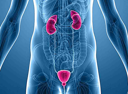
A prostate ultrasound allows for examination of the prostate gland itself, testicles, urethra, bladder, and nearby lymph nodes. The doctor examines the internal tissues of the prostate and its protective capsule, measures the organ's dimensions, and determines its structural features.
Using echo imaging, suspicious areas are identified, their size, and the clarity of their boundaries with surrounding healthy tissue are determined. If problems with urination are present, the specialist also examines the bladder, assessing the amount of fluid remaining after emptying.