Ultrasound during pregnancy
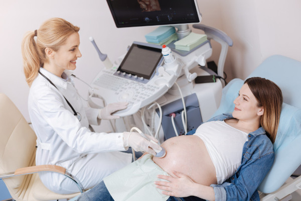
To identify the most important anatomical and functional features of the fetus, the expectant mother is prescribed one of the most informative and safe diagnostic procedures—ultrasound. Modern equipment and the experience of doctors provide reliable data, helping to promptly detect pathologies or ensure that the pregnancy is proceeding without complications.

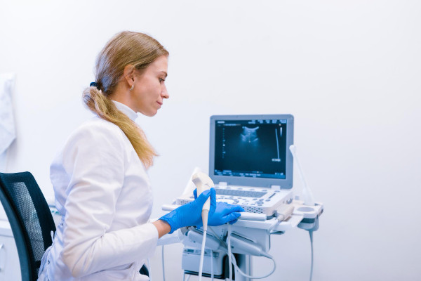
specialists

equipment

treatment


Types of ultrasound during pregnancy
Doctors monitor the pregnancy process using various types of ultrasound examinations, divided into mandatory screenings and additional selective tests. Today, ultrasound is included in the standard list of services provided to every pregnant woman in medical institutions of the country.
The main objective of mandatory screening is to identify patients with complicated pregnancy or fetal pathologies that require further in-depth examination, including the use of invasive techniques.
The purpose of any screening ultrasound is as follows:
- Determining the current gestational age
- Assessing the compliance of the fetal size with established norms
- Exclusion of hereditary defects in fetal development
- Examination of the condition of the placenta, umbilical cord and the amount of amniotic fluid
Screening is divided into stages depending on the time that has passed since conception:
- The first stage (I trimester) - is performed at 11-14 weeks
- The second stage (II trimester) - at 18–21 weeks
- The third stage (III trimester) — at 30–34 weeks
Additional (selective) ultrasound examinations are prescribed by the doctor individually when certain indications are identified, regardless of the current pregnancy period. Their appointment is due to the presence of specific risks or features of the pregnancy, such as the threat of termination, ectopic pregnancy, multiple pregnancy, abnormal position of the placenta, oligohydramnios or polyhydramnios, suspected fetal death, intrauterine hypoxia, developmental delay and other factors.
Based on the volume of information obtained and the type of image, the following types of ultrasound are distinguished:
- Two-dimensional (2D) ultrasound is a classic study that produces a familiar black and white picture in a flat form
- Three-dimensional (3D) ultrasound is a three-dimensional image of the fetus, creating a detailed portrait of the child and simplifying the diagnosis of possible developmental defects
- Four-dimensional (4D) ultrasound is similar to two-dimensional, but allows you to see the movement of the fetus in real time
- Fetometry is a procedure for measuring key parameters of the fetus (head size, chest cells, limb length, etc.) to assess the correctness of its growth and development.
- Dopplerography is a special study of blood vessels that allows you to evaluate blood circulation in the mother-placenta-fetus system, prescribed after the 20th week of pregnancy.
- Cardiotocography (CTG) - recording the fetal heartbeat and uterine contractions, used starting from the 26th week and during the labor period.
- Echocardiography is a separate diagnosis of the fetal heart, used if congenital defects of the organ or arrhythmia are suspected.
- Cervicometry - monitoring the condition of the cervix, allowing you to prevent premature birth.
The number and types of examinations are determined by the doctor based on the course of the patient's pregnancy.
First Trimester Ultrasound
In early pregnancy, ultrasound is used twice: first to confirm conception, then as part of standard first trimester screening. Transvaginal ultrasound confirms pregnancy as early as four to five weeks after the last menstrual period, while transabdominal ultrasound provides results around six to eight weeks.
An initial routine ultrasound is performed around 11 to 14 weeks. Before this time, it is impossible to thoroughly examine the embryo using this method. Along with the first trimester ultrasound, a biochemical blood test is taken to determine levels of specific substances—beta-hCG and the PAPP-A protein. These substances indicate the potential risk of severe chromosomal abnormalities in the fetus, such as Down, Edwards, Patau, and Turner syndromes.
Measurements of the nuchal translucency and nasal bone length play a key role in assessing congenital abnormalities. By combining the results of the ultrasound and biochemical test, the doctor calculates the likelihood of having a child with serious genetic disorders.
Ultrasound in the Second Trimester
The main goal of an ultrasound at this stage is to detect serious congenital defects in the fetus early. During the examination, the doctor determines the fetus's position and carefully measures the baby's key body parameters, comparing them with the expected size for this stage of pregnancy.
It is during the second trimester that the baby's sex can be determined with certainty. In a multiple pregnancy, the doctor checks the fetal positioning, ruling out dangerous options such as conjoined twins.
In addition to the ultrasound, a biochemical blood test (the "triple test") is performed in the second trimester, which involves measuring the concentration of alpha-fetoprotein, hCG, and free estriol. Abnormal test results, coupled with questionable ultrasound findings, may indicate fetal chromosomal abnormalities, which will require further invasive testing.
If a serious congenital abnormality or hereditary disease is confirmed, a panel of doctors will jointly decide whether to continue the pregnancy. The final decision rests with the woman and her family.
Third Trimester Ultrasound
The last mandatory ultrasound examination for pregnant women occurs between weeks 30 and 34. During this period, doctors seek to identify late, slowly developing anomalies and assess the fetus's readiness for the upcoming birth.
Specialists determine the baby's position, compare its size with current norms, check the functioning of internal organs, movement activity, and the condition of the most important elements of pregnancy—the placenta and amniotic fluid.
Most often, a third-trimester ultrasound is supplemented by Doppler ultrasound of the vessels connecting the uterus and placenta. This approach helps ensure that the baby is not experiencing oxygen deprivation and is ready to fully develop.
General information
Indications and contraindications for the procedure
Every expectant mother wants to be sure that her baby is safe, and an ultrasound is the best way to do this. Problems can be noticed early and, if possible, resolved or the patient's worries can be alleviated by confirming that both she and the baby are fine.
Indications for an Ultrasound
An ultrasound examination during pregnancy is prescribed to:
- Confirm pregnancy and determine whether the fetus is developing in the uterus
- See how the baby is growing and whether its size corresponds to the expected period
- Determine the exact number of babies if there appear to be two or more
- Find and correct problems with the location of the placenta if it is low or blocking the opening of the uterus
- Understand whether the baby is getting enough oxygen and nutrients by assessing blood flow in the umbilical cord and placenta
- Rule out serious fetal diseases if malformations are suspected
- Find out the number of Amniotic fluid
- Monitor for any threats of miscarriage or premature labor
- Check the condition of the cervix if there is a risk of premature dilation
The main thing is to understand that an ultrasound should not be done casually, but only when indicated.
Contraindications and limitations
Ultrasound is considered safe for pregnant women, but even it has some limitations:
- It is best to avoid unnecessary radiation, as too much can have an adverse effect on the fetus and the expectant mother's body. However, a reasonable number of ultrasounds are completely harmless.
- For expectant mothers suffering from mental disorders or severe anxiety about procedures, the doctor may temporarily postpone the ultrasound to reduce stress.
- There is a limitation on the quality of equipment: outdated devices can produce unreliable results, so only modern equipment should be trusted.
- The most important rule is that ultrasounds should be performed as prescribed by a doctor.


Pregnancy ultrasound prices in Moscow
The cost of an ultrasound for an expectant mother depends on several factors: the clinic's level and technical equipment, the doctor's qualifications, and the convenience and availability of the appointment. Modern equipment with 3D and 4D imaging capabilities increases the cost of the service, but guarantees high-quality and accurate results.
A fee-based ultrasound during pregnancy in Moscow costs approximately 2,000 to 8,000 rubles. If you're looking for a medical center where you can have a paid ultrasound in comfortable conditions without wasting precious time and receive reliable, comprehensive results, we recommend scheduling an appointment at the K+31 clinic in Moscow. You can find out the cost by phone, from the price list on our website, or during an in-person appointment with the doctor. The latter option will ensure the most accurate price.

Our doctors


This award is given to clinics with the highest ratings according to user ratings, a large number of requests from this site, and in the absence of critical violations.

This award is given to clinics with the highest ratings according to user ratings. It means that the place is known, loved, and definitely worth visiting.

The ProDoctors portal collected 500 thousand reviews, compiled a rating of doctors based on them and awarded the best. We are proud that our doctors are among those awarded.
Make an appointment at a convenient time on the nearest date
Price
Other services

Appointment to the doctor
Reviews


Our clinics

Application “Personal Account K+31”


Timing of fetal ultrasound
The development of an unborn child in the womb is a complex and ongoing process that requires regular monitoring. According to recommendations from the Russian Ministry of Health, every woman should undergo three mandatory ultrasound examinations during a normal pregnancy. If complications or signs of abnormalities arise, the gynecologist managing the pregnancy increases the number of examinations.
The most appropriate periods for ultrasound examinations are the middle of the first, second, and third trimesters, that is, approximately at the twelfth, twenty-second, and thirty-second weeks of gestation. This timing allows doctors to consistently monitor the growth and development of the fetus, identifying any abnormalities early.