Treatment of hemangioma in newborns
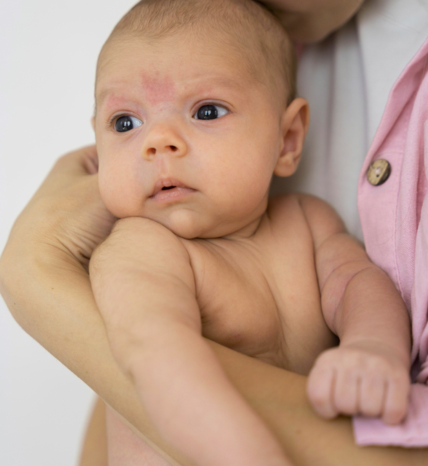
Hemangiomas in newborns are not a cause for panic, but they are also not something to ignore. These vascular growths are common, occurring in approximately 5-10% of infants. They may appear as a harmless red spot, but sometimes they grow rapidly, change the skin, and interfere with vision or breathing. It is important for parents to understand how infant hemangiomas develop and seek medical attention promptly.
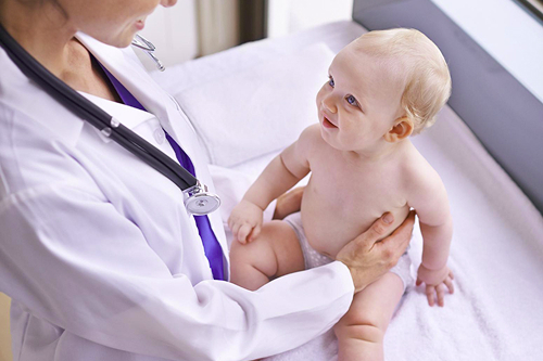
At the K+31 Clinic, hemangiomas are treated using modern methods—gently, painlessly, and with outcome monitoring at every stage.

specialists

equipment

treatment


Causes of hemangiomas
The exact cause of vascular tumors is not fully understood. It is known that hemangiomas are more common in girls, premature babies, and those with difficult births. Scientists associate their development with a temporary disruption in the development of the fetal vascular system in the first trimester of pregnancy.
Indirect risk factors may include:
- Maternal viral infections during pregnancy
- Chronic fetal hypoxia
- Maternal age over 35
- Multiple pregnancies
- Hormonal imbalances
However, hemangiomas in newborns are not associated with genetic diseases. They are spontaneous vascular formations that can be successfully managed in most cases.
Types of hemangiomas
The type of hemangioma determines not only its appearance but also its behavior—growth rate, tendency to regress, and the choice of treatment method. The doctor always evaluates the depth of skin and underlying tissue involvement and the risk of organ dysfunction or cosmetic defects.
There are several main types of vascular tumors based on structure and location:
Capillary (simple)
This is the most common form in infants, accounting for approximately 80% of all cases. It is localized in the superficial layers of the skin, most often on the face, head, or trunk. It appears as a flat or slightly raised, bright red spot with clear borders. When pressed, it fades and then refills with blood.
A capillary hemangioma typically grows during the first 6-8 months of a child's life, after which it stabilizes and gradually shrinks. The prognosis is favorable—in most cases, the lesion resolves spontaneously without treatment. However, if the spot is located near the eye, mouth, or in skin folds, a doctor may recommend medication or laser treatment to prevent irritation and trauma.
Cavernous
This form is located deeper—under the skin or in the subcutaneous tissue. The tumor consists of several large vascular cavities (caverns) filled with blood. The formation is soft and elastic to the touch and may enlarge when the child cries or coughs. The color ranges from bluish-red to purple.
Cavernous hemangiomas are most often found on the head, neck, back, and extremities. The danger lies in the possibility of vascular damage and bleeding. If the tumor is growing rapidly or is located in functionally significant areas, treatment is administered—usually medication with beta-blockers or a combination of medication and laser treatment.
Combined
The combined form combines features of both capillary and cavernous hemangiomas: part of the lesion is located on the skin's surface, while the other part is located deeper within the tissue. Externally, it appears as a spot with areas of hardening, sometimes irregular in shape.
These tumors can vary in color from light pink to dark red, have an uneven structure, and require more complex diagnostics, such as ultrasound or MRI. Combined hemangiomas tend to grow rapidly in the first months of life, so doctors often recommend early treatment to avoid cosmetic defects and skin deformities.
Mixed
The mixed form includes not only vascular tissue but also elements of adipose, lymphatic, or connective tissue. This rare type can have a dense consistency, uneven color, and a more complex structure.
Mixed hemangiomas require a multidisciplinary approach—examination by a vascular surgeon, a dermatologist, and, if deep, a pediatric oncologist. Treatment is individualized, often combining medication and instrumental treatments.
Additional forms and growth characteristics
Sometimes, doctors also distinguish internal hemangiomas—vascular neoplasms that develop in the liver, kidneys, or other organs. They are asymptomatic and detected incidentally during ultrasound.
By growth pattern, hemangiomas are classified as:
- Proliferating—actively increasing in size during the first months of life
- Involuting—gradually decreasing
- Involuted—completely disappearing or leaving a light spot on the skin
The prognosis and physician's approach depend on the type of hemangioma. Superficial lesions often regress without a trace, while deep lesions require monitoring and, if necessary, treatment. That's why accurate diagnosis of the tumor type at the K+31 clinic is the first and most important step toward a child's safe recovery.
Symptoms of hemangioma
The external manifestations of a hemangioma depend on its type and location. Most often, parents notice a red spot on the skin, which may be slightly raised and change color when crying or chilled. During the first months of life, the growth may grow rapidly—this is a natural phase of infantile hemangioma development. Then, by the age of one year, growth usually slows, and by school age, some tumors can shrink on their own.
Head lesions
Hemangiomas on the head are one of the most common locations. Here, they are subject to friction during child care, so it is important to ensure the skin is not injured. If the lesion is located near the eye or ear, it requires the attention of specialized specialists: an ophthalmologist and otolaryngologist.
Facial pathology
Facial vascular lesions are particularly noticeable and cause concern for parents. A hemangioma on the face requires careful attention—not only for its cosmetic appearance, but also because important blood vessels and nerves pass through this area. Modern methods allow for the removal or reduction of the growth without leaving marks or scars.
Hemangioma on the back
Such growths often do not interfere with organs and can be observed periodically. However, if the growth enlarges, becomes inflamed, or damages the skin due to friction, a doctor may recommend treatment to prevent complications.
Forms on the extremities
Hemangiomas of the arms and legs are noticeable within the first few days of life. They are rarely dangerous, but can interfere with mobility if located near a joint.
Internal organs
Sometimes, vascular growths occur not on the skin, but in internal organs such as the liver, kidneys, or brain. Such cases require careful diagnosis, as there are no external signs. They can be detected by ultrasound or MRI if the doctor suspects an internal hemangioma.

Diagnosing hemangioma
Diagnosis begins with an examination of the child by a dermatologist or surgeon. In most cases, a visual assessment is sufficient. The doctor determines the type, depth, and characteristics of the vascular formation.
To clarify the diagnosis, the following are used:
- Ultrasound with Doppler imaging – shows the structure and blood flow
- MRI or CT scan (for deep or extensive lesions)
- Dermoscopy of the skin surface
- Laboratory tests if complications are suspected
Differential diagnosis
The doctor must distinguish hemangioma from other skin and vascular pathologies: pyogenic granuloma, angioma, and vascular malformations. Unlike the latter, infantile hemangioma actively grows in the first months of life, then stabilizes and may regress. This is an important diagnostic feature.
Treatment of hemangiomas
Modern approaches to treating hemangiomas in newborns are aimed at preserving the child's health and appearance. At the K+31 clinic, doctors select an individualized approach: observation, drug therapy, or removal, depending on the location, growth rate, and age of the infant.
Conservative (non-invasive) treatment
Many hemangiomas do not require surgical intervention. If the growth is small, does not interfere with organ function, or damage the skin, the doctor may recommend monitoring the growth. Parents are instructed on how to monitor the spot and what signs require a follow-up examination.
In addition, medications are used, such as propranolol-based medications (beta blockers). These slow the growth of vascular cells and promote tumor regression.
Surgical (invasive) treatment
Surgical removal is performed in cases of rapid growth, bleeding, ulceration, or the risk of organ dysfunction. At the K+31 clinic, operations are performed delicately, under visualization guidance, with minimal tissue trauma.
Systemic treatment methods
Systemic therapy is used for multiple or extensive lesions. It aims to stabilize vascular growth and prevent complications.
Localized treatment methods
Local treatments include laser removal and cryodestruction. Lasers allow for targeted treatment of blood vessels without damaging the surrounding skin. This is a safe method that is well tolerated by infants and produces a noticeable cosmetic effect.
Other treatments
Depending on the situation, the doctor may use a combination of approaches: medication, phototherapy, and microwave therapy. The main goal is to remove or shrink the hemangioma painlessly and without leaving marks on the baby's skin.
Complications of hemangioma

Despite its benign nature, vascular lesions can cause complications, especially if they grow rapidly or if the surface is damaged.
The most common consequences are:
- Ulceration and bleeding
- Infectious inflammation
- Impaired vision, hearing, and breathing (if located in critical areas)
- Scarring of the skin after injury
At first glance, a small red spot appears harmless, but its cells are actively dividing, and the vascular network is very delicate. Even light friction from clothing, a diaper, or scratching can damage the epidermis. Infection can enter through microcracks, and a benign process turns into an inflamed, painful wound.
Ulcerations most often develop in skin folds, on the lips, chin, armpits, and groin area—areas with a moist environment and constant friction. These wounds heal slowly, cause pain to the infant, and may leave pigmented or scarred marks. In severe cases, there is a risk of secondary bacterial infection.
If the vascular lesion is located on the face, eyelids, or nose, complications can affect the sensory organs. The tissue growth obscures normal vision and compresses the ear canal or airway. In newborns, such conditions develop quickly, so any slight changes in color or volume should promptly bring the child to a doctor.

Sometimes complications are not related to tumor growth, but to attempts at self-intervention. Parents, wanting to "cauterize" the spot, use harsh ointments, alcohol-based tinctures, and compresses. Not only are these methods ineffective, but they also damage the baby's delicate skin, causing burns and extensive inflammation. Even a small lesion can leave noticeable scars that require subsequent correction.
Bleeding is also dangerous: vascular tissue is rich in capillaries, and if its integrity is compromised, even minor trauma can cause significant blood loss. This condition develops more quickly in newborns than in adults, so any bloody discharge from the surface of the tumor is a signal to seek medical attention.
In rare cases, large vascular lesions can affect the child's general condition. Due to constant blood flow in the tumor vessels, the strain on the cardiovascular system increases, resulting in decreased hemoglobin, weakness, and pale skin. Such situations require observation by a pediatrician and vascular surgeon, and sometimes hospitalization.

If the lesion remains unattended for a long time, skin deformation may occur:
- The area becomes denser
- Loses elasticity
- Indentation or unevenness appears
This is not dangerous to health, but it can become a significant cosmetic defect.
Therefore, any attempts at self-removal, the use of untested remedies, or "home remedies" are strictly contraindicated. Only a specialist can assess the depth of the lesion, determine the type of vascular structure, and choose a safe treatment method.
When visiting the K+31 clinic, a child is examined by a multidisciplinary team of doctors—a pediatrician, surgeon, and dermatologist. This approach allows us to identify complications at an early stage and choose the optimal treatment strategy: drug therapy, gentle removal, or ultrasound-guided monitoring.
Timely assistance prevents pain, bleeding, and inflammation, and most importantly, keeps the baby's skin healthy and free of traces of intervention.
General information
Prognosis and prevention
The prognosis for infantile hemangiomas is favorable. Most lesions gradually disappear by 5-7 years of age, leaving no trace. However, some require treatment to avoid cosmetic or functional problems.
There is no specific prevention. The main thing is to be monitored by a pediatrician and promptly seek medical attention if a spot or nodule appears on an infant's skin.

FAQ
Is it possible to simply wait for the hemangioma to resolve on its own?
Sometimes, yes. But only after a doctor's examination confirms the growth is safe.
Is laser treatment painful for children?
Modern lasers are painless. Local anesthesia is used if necessary.
Will removal leave marks on the skin?
In most cases, the skin recovers completely after treatment, without scarring or pigmentation.
When to see a doctor?
If the spot grows, changes color, ulcerates, or bothers the baby, see a doctor as soon as possible.
The K+31 Clinic brings together experienced specialists in the treatment of vascular pathologies in children. We approach each case individually, taking into account the child's age and health characteristics.
You can schedule a consultation with a dermatologist or pediatric surgeon at the K+31 Clinic online or by phone. Early consultation is the key to a calm life and healthy skin for your child.
Our doctors


This award is given to clinics with the highest ratings according to user ratings, a large number of requests from this site, and in the absence of critical violations.

This award is given to clinics with the highest ratings according to user ratings. It means that the place is known, loved, and definitely worth visiting.

The ProDoctors portal collected 500 thousand reviews, compiled a rating of doctors based on them and awarded the best. We are proud that our doctors are among those awarded.
Make an appointment at a convenient time on the nearest date
Price
Other services

Appointment to the doctor
Reviews


Our clinics

Application “Personal Account K+31”


What is a hemangioma?
A hemangioma is a benign vascular tumor that occurs in infants in the first weeks or months of life. It develops from cells in the vascular wall that begin actively dividing after birth. On a newborn's skin, a hemangioma most often appears as a red or burgundy spot that may protrude slightly above the surface.
While the word "tumor" sounds alarming, it's important to understand: an infant hemangioma is a benign growth that is not prone to malignant transformation. However, under certain conditions (such as rapid growth or damage to the skin surface), the growth requires monitoring and treatment.