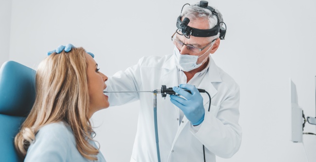
Laryngoscopy – endoscopy of the throat and larynx

specialists

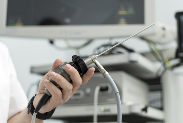
equipment

treatment
Endoscopic laryngoscopy can detect laryngeal inflammation and abscesses. It is also carried out to detect:
- Tumors (benign and malignant)
- Burns caused by thermal or chemical exposure
- Adhesions and tumors on the vocal cords
This procedure also helps to identify injuries to the vocal cords and detect the presence of foreign objects in the larynx.

Indirect laryngoscopy is usually safe and does not have strict restrictions on its use. Direct laryngoscopy has contraindications.
These include:
- Problems with the blood coagulation system
- Epileptic seizures
- Recent throat surgery
- Serious cardiovascular diseases
The procedure is not performed in the presence of these conditions, because this background increases the risk of complications.
General information about the procedure
Types of procedure
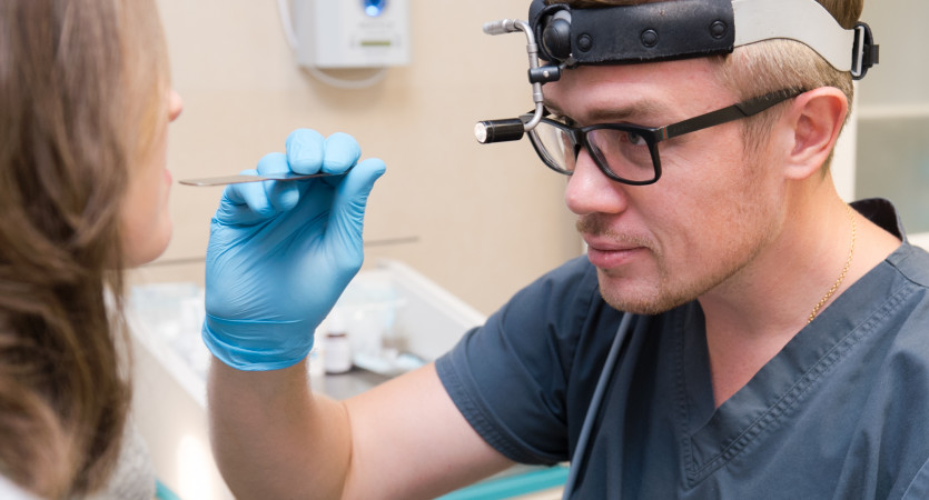
Laryngoscopy can be indirect, direct or retrograde. During indirect laryngoscopy, the doctor examines the throat using a special mirror.
Direct laryngoscopy allows for a more in-depth examination. The process uses flexible (used with local anesthesia) or rigid (requires general anesthesia) fiber laryngoscopes with an optical system.
Retrograde laryngoscopy is used to examine the nasopharynx through the nose. This is a minimally invasive procedure that is performed under local anesthesia.


K+31 photo gallery


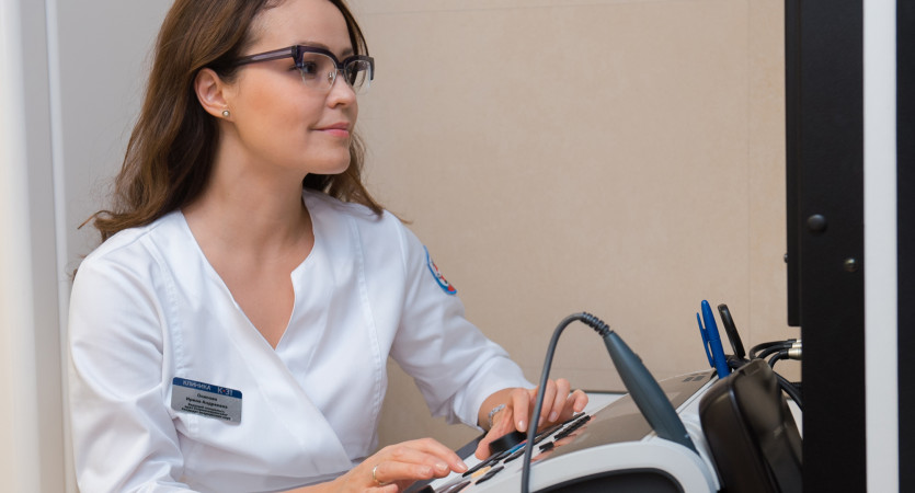


Our doctors


Make an appointment at a convenient time on the nearest date

This award is given to clinics with the highest ratings according to user ratings, a large number of requests from this site, and in the absence of critical violations.

This award is given to clinics with the highest ratings according to user ratings. It means that the place is known, loved, and definitely worth visiting.

The ProDoctors portal collected 500 thousand reviews, compiled a rating of doctors based on them and awarded the best. We are proud that our doctors are among those awarded.
Price
Other services

Appointment to the doctor


Reviews
Our clinics

Application “Personal Account K+31”


