Rhinoscopy

The purpose of rhinoscopy is to thoroughly examine the internal structures of the nose, pharynx and sinuses using specialized instruments. This diagnostic method allows the otolaryngologist to evaluate the anatomical and functional features of the nasal passages, detect the presence of pathologies or inflammatory processes and determine their nature.
Best
specialists
Expert
equipment
Advanced diagnostic
treatment
Sign up
Fill out the entry form on the page and we will call you back
Submit your application

When is rhinoscopy necessary?

The procedure is performed for frequent nosebleeds.

It is also shown when:

  • Difficulty in nasal breathing
  • Nasal congestion
  • Lack of sense of smell
  • The presence of purulent or clear discharge from the nasal cavity
  • Severe headaches

In addition, it is prescribed for identified diseases of the nasopharynx. Rhinoscopy can be anterior, middle and posterior.

When is rhinoscopy necessary?
General information about the procedure

Anterior rhinoscopy

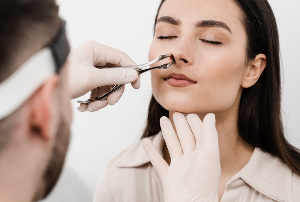
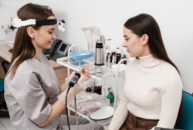
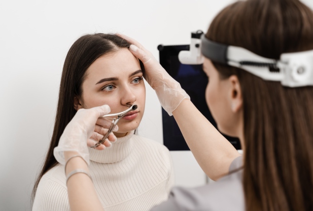
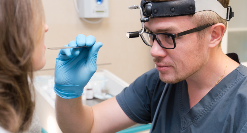
Direct or anterior rhinoscopy involves examining the inside of the nose using a special instrument - a dilator. In this case, the patient sits opposite the doctor, who, holding his head, inserts the instrument into the nasal passage. The depth of its insertion varies depending on the age of the subject and the purpose of the examination.

By changing the position of the patient's head, the doctor examines different parts of the nasal cavity. For example, when looking straight, the lower areas are accessible, and when the head is tilted back, the middle areas are accessible. Particular attention is paid to the middle meatus, since this is where important anatomical structures are located.

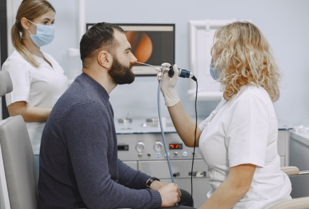
During the procedure, the specialist assesses the condition of the mucous membrane, the size of the nasal turbinates and septum, as well as the amount and nature of discharge. In some cases, an additional examination of the posterior wall of the nasopharynx is carried out - this helps to diagnose, for example, adenoiditis. At the same time, for a better view, the doctor asks the patient to make sounds or move his head.

Rhinoscopy of the nasopharynx should not cause pain. If the patient experiences discomfort, the doctor administers pain relief (local agents are used).

To assess the condition of the mucous membrane and identify pathological formations, it is additionally palpated with a special probe. Anemization of the mucous membrane with vasoconstrictor drugs improves visibility and helps diagnose rhinitis.

Inspection of the middle meatus

This rhinoscopy allows the doctor to carefully examine the middle meatus, the upper two-thirds of the septum, the openings of the maxillary and frontal sinuses, as well as the semilunar cleft. Sometimes during the procedure, an additional examination of the posterior wall of the nasopharynx is performed. To do this, the specialist uses a special expander with long levers.

During the procedure, the doctor evaluates the color and condition of the mucous membrane, the patency of the passages, the presence of curvatures or defects on the septum. He also clarifies the signs of pathological formations and the characteristics of nasal discharge.

To reduce discomfort, reduce the risk of severe swelling and thereby improve visibility of the area being examined, the doctor uses vasoconstrictor drugs.

Examination of the posterior part of the nasopharynx

Posterior rhinoscopy allows you to examine the choanae and pharyngeal openings of the auditory tubes. The doctor also examines the back of the turbinates, the septum (in the appropriate area) and the soft palate. A nasopharyngeal speculum is used for this. To access the area behind the uvula, the specialist holds the patient's tongue with a spatula. In addition, trying to provide himself with better visibility, the doctor asks the patient to breathe through his nose.

A local anesthetic spray is used to prevent discomfort. Lighting for the procedure is provided using a reflector aimed at the mirror.

Surgery through rhinoscopy

This method is an in-depth version of endoscopy. The procedure is performed under general anesthesia.

To provide free access to the target areas, the doctor removes the altered tissue. After this, the doctor uses an endoscope to carry out the necessary surgical interventions inside the nasal cavity.

Operative rhinoscopy is necessary for:

  • Removal of polyps
  • Restoration of the openings of the paranasal sinuses
  • Getting rid of fungi in the sinuses
  • Correction of anatomical structures of the nose
  • Removing foreign objects
  • Treatment of cysts and other formations

This method also allows you to accurately diagnose the presence of tumors. Therefore, it is often used for biopsy.

Safety and risks during rhinoscopy

Rhinoscopy is considered a safe procedure and does not lead to complications. However, patients with sensitive or inflamed mucous membranes often experience nosebleeds due to the mechanical impact of instruments.

The use of anesthesia carries the risk of laryngospasm, and in severe cases laryngeal edema and anaphylactic shock are observed. Therefore, before using an anesthetic, the doctor must check with the patient whether he suffers from allergies to a particular drug.

Features of nasal rhinoscopy in children

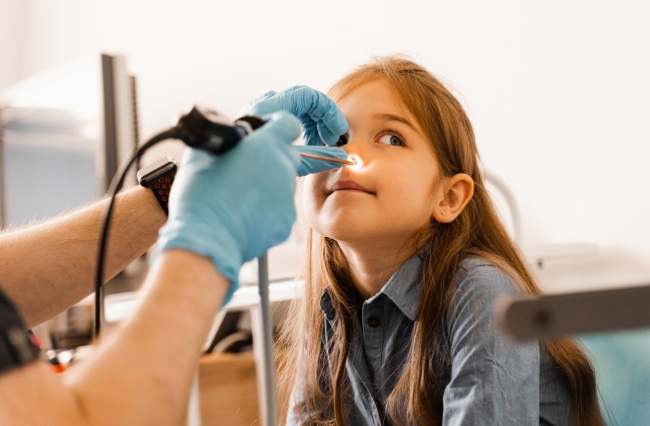
Infants and preschool children often react negatively to medical procedures. Therefore, specialists working in private clinics try to perform rhinoscopy on a child as quickly and as gently as possible. In most cases, doctors abandon the use of nasal dilators in favor of smaller instruments, such as ear specula.

In this case, the doctor limits himself to a visual examination, slightly lifting the tip of the child’s nose. If a dilator is still needed, the mucous membrane is pre-treated with an anesthetic spray.

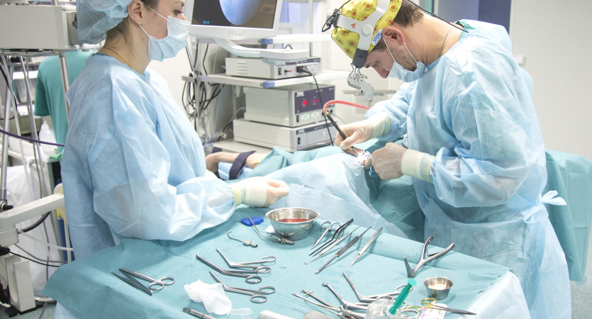
In more complex situations, when a thorough examination or surgical procedures are required, rhinoscopy in children is performed under general anesthesia. This allows you to simultaneously examine the nasal cavity and carry out the necessary manipulations, for example, take biomaterial for analysis.

Our doctors
Sadikov
Ilya Sergeyevich
Head of the Clinic for Otorhinolaryngology, Otorhinolaryngologist
Tetzoeva
Zalina Muratovna
Head of the department of otorhinolaryngology, otorhinolaryngologist
Lapshina
Anastasia Andreevna
Otolaryngologist, audiologist
Lopatin
Andrew Stanislavovich
Chief Specialist in Otorhinolaryngology
Portnyagina
Maria Pavlovna
Audiologist-otorhinolaryngologist
Kaspranskaya
Galina Rustemovna
Otorhinolaryngologist, otoneurologist, audiologist
Budeikina
Liliya Sergeevna
Otorhinolaryngologist, phoniatrist
All specialists
Make an appointment at a convenient time on the nearest date

Price

Primary appointment with an otorhinolaryngologist
from 4 950 ₽
Repeated appointment with an otorhinolaryngologist
from 4 400 ₽

Appointment to the doctor

Fill out the form, our managers will contact you within 15 minutes
Почему К+31?
К + 31 — full-cycle multidisciplinary medical centers, including the possibility of providing medical services of European quality level.
К + 31 — are leading doctors and diagnostics using high-tech equipment from world manufacturers (Karl Storz, Olympus, Siemens, Toshiba, Bausch&Lomb, Technolas, Zeiss, Topcon).
К + 31 — is ethical. The staff of K+31 clinics maintain open relationships with patients and partners. An individual approach to each patient is the basis of our service standards.
К + 31 — is modernity. On call 24/7: call center operators will answer your questions at any time and book you an appointment with doctors. Contact us by phone, through the feedback form on the website and WhatsApp.
Our clinics
K+31 on Lobachevskogo

st. Lobachevskogo, 42/4

+7 499 999-31-31

Subway
1
11
Prospect Vernadsky Station
By a car
Lobachevsky, we pass the first barrier (security post of the City Clinical Hospital No. 31), turn right at the second barrier (security post K+31)
Parking pass
Opening hours
Mon-Fri: 08:00 – 21:00
Saturday: 09:00 – 19:00
Sunday: 09:00 – 18:00
K+31 Petrovskie Vorota

1st Kolobovsky lane, 4

74999993131

Subway
9
Tsvetnoy Bulvar
10
Trubnaya
By a car
Moving along Petrovsky Boulevard, turn onto st. Petrovka, right after - on the 1st Kolobovsky per. Municipal parking
Opening hours
Mon-Fri: 08:00 – 21:00
Sat-Sun: 09:00 – 19:00
K+31 West

Orshanskaya, 16/2; Ak. Pavlova, 22

74999993131

Subway
3
Molodezhnaya
By a car
Moving along Orshanskaya street, we turn to the barrier with the guard post K+31. You do not need to order a pass, they will open it for you
Opening hours
Mon-Fri: 08:00 – 21:00
Sat-Sun: 09:00 – 18:00
Didn't find the service you were looking for?