Bile duct cancer

Bile duct cancer

Bile duct cancer (also known as cholangiocarcinoma) is a rare malignant tumor that develops from cells internal lining of the bile ducts. The disease can be asymptomatic, and therefore the treatment of bile duct cancer often begins in the later stages. In addition, malignant tumors in this place are very "uncomfortable", and their surgical removal is difficult.

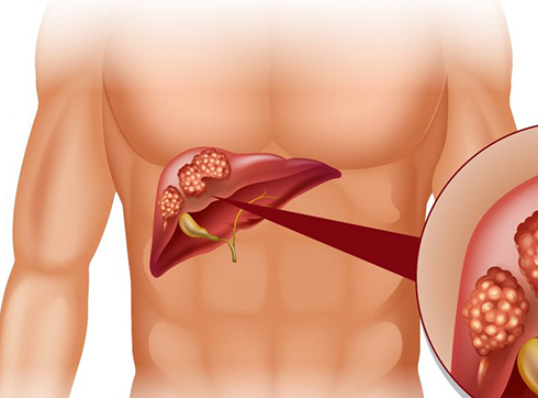
The liver is pierced by intrahepatic bile ducts, which unite at the exit from the liver into the common hepatic duct. The gallbladder is located under the liver. The bile duct comes out of it, which combines with the hepatic duct into the common bile duct (choledochus). The latter then unites with the pancreatic duct and opens into the duodenum. Tumors can develop anywhere in these intrahepatic and extrahepatic bile ducts.

Types of bile duct cancer

According to the histological structure of the tumor in the bile ducts, i.e. According to the type of cells in which it was formed, the following types of cancer are distinguished:

  • Adenocarcinoma is a malignant neoplasm that develops from the glandular cells of the inner lining of the ducts - this is the most common cancer of the bile ducts;
  • squamous cell carcinoma - from squamous epithelial cells;
  • lymphoma - from cells of lymphoid tissue;
  • sarcoma - from connective tissue cells.

A very common form of cancer of the liver or bile ducts is metastatic cancer, which has arisen from tumor cells (metastases) that have got here through the bloodstream or lymph from other organs. In this case, differential diagnosis is needed to determine the primary tumor and, accordingly, cancer treatment methods are selected, depending on its original nature. In our clinic, the root cause of bile duct cancer is precisely determined, which allows choosing the most effective treatment.

According to the localization of the tumor in the biliary tract, they are distinguished:

  • Intrahepatic cholangiocarcinoma - this is slightly less common and can be confused with liver cancer (hepatocellular carcinoma).
  • Extrahepatic cholangiocarcinoma, which is divided into the following types:
    • Proximal, or hilus, cholangiocarcinoma - it is localized to the confluence of the bile and hepatic ducts, i.e. closer to the liver. This type of bile duct cancer is more common - in more than half of cases.
    • Distal cholangiocarcinoma - it is located further from the liver and closer to the place where the common bile duct enters the duodenum. It is sometimes difficult to distinguish from pancreatic cancer.

In terms of surgical options for cancer treatment, neoplasms are divided into resectable tumors (they can be removed with a surgical operation) and unresectable (they are in hard-to-reach places or have spread greatly).

Causes of bile duct cancer

The root cause of any malignant neoplasms is mutations in normal cells that cause these cells to divide uncontrollably. But the causes of these mutations are not fully understood. However, there are risk factors that contribute to a higher likelihood of mutations and tumor development in the bile ducts.

Such risk factors include:

  • Male gender. Bile duct cancer is more common in men.
  • Age. With age, the number of mutations in cells accumulates, so the likelihood of carcinogenic mutations increases.
  • Inflammation. The inflammatory process increases the risk of cancer cells. In the case of the bile ducts, the primary sclerosing cholangitis disease is dangerous. It is rare and increases the chance of developing cancer by 5-10%.
  • Congenital anomalies of the biliary tract. Anomalies such as choledochal cysts, which some people have from birth, may not manifest themselves for a very long time. But they increase the risk of bile duct cancer by 30%. Hereditary pathological anomaly Caroli syndrome can also lead to the development of malignant neoplasms.
  • Parasites. Chronic parasitic diseases such as opisthorchiasis (which can be contracted through fish) are a risk factor for bile duct cancer.
  • Some diseases. For example, gallstone disease can cause inflammation and, consequently, cancer. Normal liver tissue with cirrhosis turns into scar tissue, which contributes to the occurrence of tumors. Patients with chronic ulcerative colitis often have sclerosing cholangitis. Chronic hepatitis B and C also increase the likelihood of developing bile duct cancer.

Risk factors also include alcoholism and smoking, overweight and diabetes, non-alcoholic fatty liver disease, chronic pancreatitis, exposure to certain substances such as radon, dioxins, asbestos, etc.

Symptoms of bile duct cancer

Bile duct cancer practically does not manifest itself in the early stages of development. Symptoms appear when the tumor already interferes with the passage and outflow of bile through the ducts. Even later, symptoms appear if it is cancer of the intrahepatic bile ducts.

Universal and most common symptoms include:

  • Mechanical jaundice. It manifests itself in dark yellow coloration of the skin, sclera of the eyes, mucous membranes. The reason for the yellow color is the entry of bilirubin into the blood due to the fact that the outflow of bile is impaired.
  • Skin itch. He is often out of control. This is also a manifestation of an increased level of bilirubin in the blood.
  • Darkening of urine and lightening of feces. This is due to the fact that bile does not enter the intestines, and the body tries to excrete excess bilirubin through the kidneys and urine.
  • Pain in the upper abdomen on the right side.
  • Lack of appetite or decreased appetite.
  • Nausea, maybe vomiting.
  • Weight loss.
  • Fever.

The clinical picture of bile duct cancer is complemented by the fact that the body is poisoned by bile formation products in the liver, which primarily destroy its cells. Blood clotting is impaired.

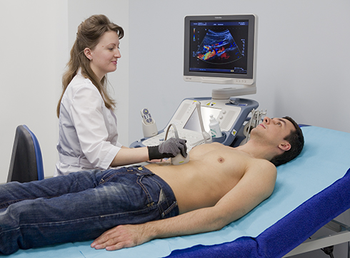
But the presence of these symptoms does not mean oncology. Bile duct cancer is rare, and the symptoms described above are more likely to indicate diseases of the gallbladder, liver, or pancreas. But in order to exclude oncology, and in case of cancer, to start its treatment as soon as possible, you need to be examined by a doctor. You can do this in our clinic by calling or signing up for an online consultation.

Stages of bile duct cancer

The most common staging system for bile duct cancer (like any other cancer) is the TNM system. It is named after three indicators: T - the size of the primary tumor, N - the number of affected regional lymph nodes, M - the presence or absence of distant metastases. Each indicator is accompanied by a number indicating the degree of expression of this feature. In the process of development, the tumor in the biliary tract increases in size, can capture neighboring tissues, its metastases affect the nearest lymph nodes, and over time, metastases reach more distant places, where they settle and form new foci.

A more familiar, simplified cancer staging system is as follows:

  • Stage 1 - the tumor has not yet spread to neighboring tissues, but is localized only within the bile duct;
  • Stage 2 - the lesion begins to invade tissues adjacent to the bile duct (liver, blood vessels, adipose tissue - depends on the location of the tumor)
  • Stage 3 - the degree of spread of the tumor to neighboring tissues increases and in parallel the cancer captures the lymph nodes
  • Stage 4 - metastases have appeared in other organs - distant parts of the liver, lungs, bones, peritoneum, etc.

There are slightly different staging classifications separately for intrahepatic bile duct cancer and extrahepatic bile duct cancer, which take into account the location of the tumor and adjacent structures, organs, and tissues.

Bile duct cancer

Diagnosis

There are no screening tests for the mass examination of patients for the presence of malignant tumors in the bile ducts. Detection of cancer usually occurs after patients consult a doctor with pain, when the disease is already in the later stages of development, and the tumor has captured neighboring tissues and metastasized. In this case, the following methods are used to diagnose:

  • Ultrasound. With the help of an ultrasound examination, it is possible for the patient to safely detect tumor formations in the bile ducts and other organs of the abdominal cavity. In this case, one of two options for ultrasound is assigned:
    • transabdominal ultrasound is the usual type of ultrasound, familiar to everyone, which is carried out through the wall of the abdomen;
    • endoscopic ultrasound is an endoscopic examination in which an endoscope with a probe at the end is inserted into the duodenum.
    Ultrasound allows you to determine the location of the tumor in the biliary tract, its size and extent of spread.
  • Cholangiography is an X-ray diagnostic method when the bile ducts are filled with a special solution, to make them visible and clearly visible in the picture. This examination allows you to determine the localization of the tumor. Cholangiography can be percutaneous and performed with a needle or endoscopic (ERCP - endoscopic retrograde cholangiopancreatography) - using an endoscope and a thin catheter that is inserted into the bile duct. This procedure also allows you to take cancer cells for biopsy.
  • A biopsy is a histological examination that allows you to accurately determine the nature of the tumor in the biliary tract and make a reliable diagnosis. Cell sampling is carried out in different ways - using cholangiography, using a needle under ultrasound or CT control, etc.
  • Computed tomography and magnetic resonance imaging - these diagnostic methods help to assess the location of the tumor, its size, the degree of spread to the surrounding tissues, the nature of the tumor (benign or malignant), and to assess the possibility of surgical intervention.
  • Laparoscopy is a surgical procedure in which a laparoscope is inserted through an incision in the abdomen to view the abdominal cavity. This way you can determine the stage of bile duct cancer, detect other pathologies and tissue lesions and, if necessary, perform a biopsy.

Suspicion of bile duct cancer can be checked using a biochemical blood test if an increase in the concentration of all fractions of bilirubin is observed.

Cancer of the proximal extrahepatic bile ducts is named after the doctor who first described this neoplasm in detail - Klatskin's tumor. The presence of this disease complicates the diagnosis. By the time jaundice appears, the tumor has usually grown into the ducts of the liver. It can affect the hepatic arteries and branches of the portal vein, and this makes it impossible to use surgical methods for treating cancer.

To make a diagnosis, if a tumor of the liver, bile ducts and pancreas is suspected, the patient is sent for laboratory blood and urine tests. Among the methods of hardware diagnostics, ultrasound of the abdominal organs, esophagogastroduodenoscopy, X-ray of the chest organs, CT, MRI, angiography, and laparoscopy showed high efficiency. This is quite enough to identify the tumor, accurately determine its location and size, and also find out which organs metastases have spread to, if any. Based on laboratory tests and hardware examination, the doctor receives data on the resectability of the tumor, that is, on the possibility of radical surgery.

Bile duct cancer treatment

The choice of cancer treatment depends on the size of the tumor, its stage, location. And also from the state of the liver and the patient's body, from the presence of other concomitant diseases.

Surgical treatment

The most effective treatment for bile duct cancer is removal of the tumor. But such a procedure is possible only in the early stages.

If the neoplasms are located in the intrahepatic ducts, liver resection can be extensive (the entire lobe of the liver is removed) or economical. After the operation, the organ continues to function normally.

If the tumors are located in the extrahepatic ducts, a much more complicated operation is ahead, during which sections of not only the ducts, but also neighboring organs (liver, gallbladder, pancreas, duodenum), as well as lymph nodes can be removed.

If there is cirrhosis, and the tumor is one and small, then the oncologist surgeon can perform liver transplantation, since cirrhosis greatly impairs the functions of this organ.

Radiation therapy

Radiotherapy is given before surgery to reduce the size of the tumor, or after it to prevent recurrence. In the advanced stages of cancer, radiation therapy is used as a palliative treatment to reduce symptoms and alleviate the patient's condition.

Chemotherapy for cancer

Chemotherapy for bile duct cancer, like radiation therapy, is used before or after cancer surgery for the same purposes. Chemotherapy as an independent method of treatment is palliative in nature and is prescribed for patients with advanced cancer. It can complement liver transplantation. Chemotherapy is often combined with radiation therapy - chemoradiotherapy.

Prognosis of survival in bile duct cancer

Due to the lack of screening tests, the disease is asymptomatic until the last stages, and tumors are often located in hard-to-reach places, the five-year survival rate for bile duct cancer is very low - an average of 8%. At the first stage of the disease, it is less than 25%. When a tumor in the bile ducts has spread to neighboring tissues and lymph nodes, this figure drops to 6%.

Bile duct cancer prevention

It is impossible to influence all risk factors. But some of them are in the power of man. For prevention, it is worth giving up bad habits, maintaining a normal weight, eating right and leading a healthy lifestyle, getting vaccinated against hepatitis B, and practicing safe sex. And in the presence of hepatitis or other diseases from the risk zone, you need to undergo regular examinations.

Preventive measures for early detection of a tumor include:

  • examination by a general practitioner and oncologist at least once a year;
  • CT or MRI (optional, if cancer is suspected based on test results);
  • testing for oncomarkers CEA, SA-19.9, AFP;
  • cholangiography (in the presence of previously installed drains);
  • Ultrasound of the abdominal organs twice a year.

Our clinic provides diagnostics and treatment of stomach cancer, breast cancer, lung cancer, colon cancer, pancreatic cancer, esophageal cancer, uterine cancer, ovarian cancer, rectal cancer, bladder cancer, kidney cancer and other cancers. diseases. Give us a call to make an appointment or leave a request on the website, and we will contact you as soon as possible.

Service record


Specialists

All specialists
Merkulov
Igor Alexandrovich

Deputy chief physician for oncology, oncologist

Doctor of Sciences, PhD


Academician, professor, Doctor of Sciences, PhD

Petrov
Dmitry Yurevich

Deputy chief physician for oncology, surgeon

PhD, Docent

Ershova
Ksenia Igorevna

Head of department, oncologist

PhD

Rasner
Pavel Ilyich

Consultant in urology, urologist

Doctor of Sciences, PhD, professor

Malygin
Sergey Evgenyevich

Oncologist-mammologist, surgeon

Pshikhachev
Ahmed Mukhamedovich

Urologist, Oncologist

Doctor of Sciences, PhD

Shevchuk
Alexei Sergeyevich

Oncogynecology consultant, obstetrician-gynecologist

PhD

Chichkanova
Tatyana Vladimirovna

Oncologist-mammologist, radiologist

Katz
Ksenia Vladimirovna

Dermatovenerologist, oncologist

Rakova
Elena Sergeevna

Oncologist, chemotherapist

Udin
Oleg Ivanovich

Deputy chief physician for surgery, surgeon

PhD

Kogonia
Lali Mikhailovna

Chemotherapist

Doctor of Sciences, PhD

Ushenina
Maria Valerievna

Oncologist-chemotherapist

PhD

Stepura
Yulia Evgenievna

Head of the department of antitumor drug therapy, oncologist, chemotherapist

Volkova
Daria Mikhailovna

Head of the radiation therapy department

Gomov
Mikhail Alexandrovich

Consultant in oncogynecology, obstetrician-gynecologist

Grishin
Igor Igorevich

Obstetrician-gynecologist

Doctor of Sciences, PhD, professor

Achba
Maya Otarovna

Radiologist, ultrasound diagnostician, oncologist-mammologist