Ultrasound or mammography: how to choose the right breast examination method

When it comes to breast health, women often ask the same question: "Which is better — ultrasound or mammography?" The correct answer is not "which is better", but "what and when". These two methods solve different problems, and together they make up a full—fledged diagnostic shield, whereas individually each of them has its own blind spots.

Best specialists
Best
specialists
Expert equipment
Expert
equipment
Advanced diagnostic treatment
Advanced diagnostic
treatment
Download the application “Personal Account K+31”
Everything about your health in one click!
Subscribe to social networks K+31
To keep up to date with current offers and receive useful advice about your health.

The principle of operation of each method
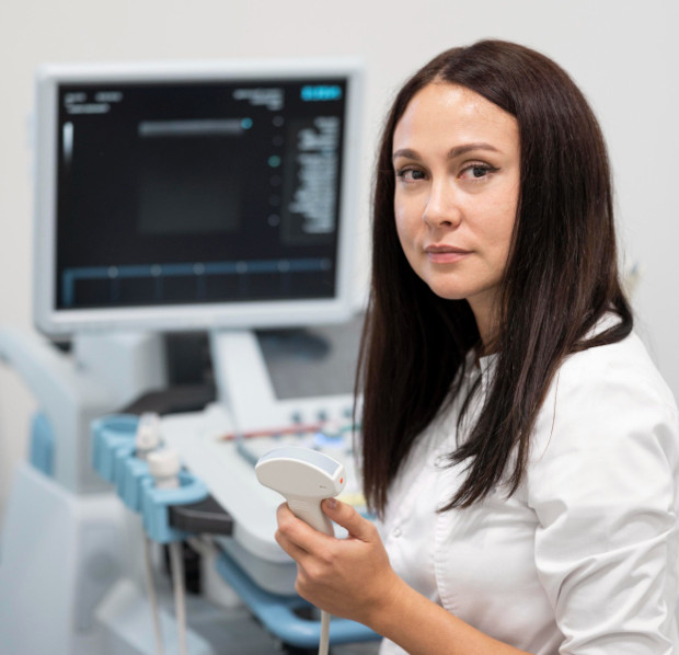
Mammography

Mammography

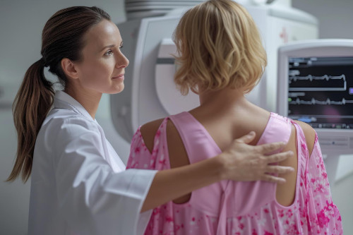
X-ray examination of the mammary glands: the tissue is compressed between two plates, images are taken in two projections (straight and oblique). Modern digital mammographs (including tomosynthesis — "3D mammography") provide a layered image and significantly reduce the proportion of false positive results.

Strengths:
  • Detection of microcalcinates, tiny calcium deposits that are one of the early markers of cancer in situ (DCIS)
  • Detection of structural rearrangements of the gland tissue
  • The ability to compare images in dynamics from year to year
  • Information content is higher with the fatty type of gland (predominates after 40-45 years)
Restrictions:
  • Radiation dose (minimal, but existing) — excludes regular use in young women and during pregnancy
  • Decreased sensitivity in dense glandular tissue — in young women, mammography sensitivity drops to 50-60%
  • Pain during gland compression (for some patients)
  • It is not suitable for the examination of implants without a special protocol

Breast ultrasound

Breast ultrasound

The ultrasonic sensor scans the tissue without compression and without radiation exposure. Modern high-frequency sensors (15-22 MHz) provide sufficient resolution to detect formations from 3-5 mm. Strengths:
  • Absolute safety — allowed during pregnancy, lactation, and in adolescents
  • High information content with dense glandular tissue — it is the ultrasound that "sees" what the mammograph "loses" in young women
  • Distinguishing between cysts and solid formations (accuracy up to 98-100%)
  • Assessment of blood flow in education (Doppler) is an important criterion of malignancy
  • Navigation during biopsy — puncture under ultrasound control in real time
  • Assessment of lymph nodes in the axillary zone
  • There are no restrictions on the frequency of the event
Restrictions:
  • It does not detect microcalcinates, the main early sign of cancer in situ
  • Decreased sensitivity in case of fatty gland involution
  • Operator dependence: the result is largely determined by the doctor's qualifications
  • The impossibility of a full assessment in dynamics (unlike snapshots)

Comparison table

Parameter Breast ultrasound Mammography
The principle Ultrasound X-ray radiation
Radiation load No Minimum (~0.4 mSv)
Dense glandular tissue High precision Reduced
Fat involution Reduced High precision
Microcalcinates Does not detect Reveals well
Cysts Accuracy ~100% Limited
Biopsy under control Yes Yes (stereotaxically)
Pregnancy/lactation Yes No
Pain during examination No Possible
Implants Without restrictions Special protocol
Screening (50-70 years old) Addition The "Gold Standard"
doctor k31

To whom and when is what prescribed?

Up to 35-40 years old is the primary choice: ultrasound

In young women, dense glandular tissue prevails, in which the mammograph "goes blind". Ultrasound at this age is much more informative. Routine preventive examination is annual ultrasound of the mammary glands.

40-50 years old — both methods in tandem

The glandular tissue is changing: glandular is replaced by fatty, and the sensitivity of mammography is increasing. Recommended:

  • Ultrasound scan — 1 time per year
  • Mammography — once every 1-2 years

50+ years old — mammography as a basis, ultrasound as a supplement

The fatty gland type makes mammography the "gold standard" of screening. WHO, EUSOMA and the Ministry of Health of the Russian Federation recommend mammographic screening specifically for this age group.

Mammography is performed once a year; Ultrasound is added when:

  • detection of compaction
  • ambiguous mammographic picture
  • dense gland according to mammography (BIRADS C/D)

Special situations — always ultrasound (± MRI)

  • Pregnancy and lactation
  • Breast implants
  • Severe breast pain
  • Mastitis, breast abscess
  • Puncture of the formation or biopsy
  • Women with BRCA1/BRCA2 mutation (optional MRI)

General information

Sensitivity of methods: evidence-based medicine figures

The situation ULTRASOUND Mammography ULTRASOUND + mammography
Women under 40 (dense gland) up to 95% 50–60% up to 98%
Women 40-50 years old ~80–85% ~75–85% ~95%
Women 50+ (adipose gland) ~75% ~90–97% ~98%
Cysts ~99–100% ~70–80%
Cancer in situ (DCIS with calcinates) ~30–40% ~80–90%

The data confirm that the combination of methods increases the detection of breast cancer by 9-15% compared with each method individually.
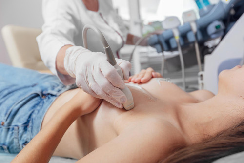
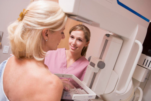
Sensitivity of methods: evidence-based medicine figures

Ultrasound and mammography at K+31

In the K+31 clinic, both methods are performed using modern equipment.:

  • Senograph Crystal Nova (GE)Digital Mammograph — reduced radiation exposure, high resolution, targeted magnification
  • Expert-class ultrasound devices with high-frequency sensors and Dopplerography

K+31 mammologists combine specialization in radiation diagnostics and oncomammology, which means a single interpretation. all the results and the possibility of immediate consultation with an oncologist in case of suspected pathology.

When are both methods needed at the same time?

  1. BIRADS 0 on mammography — ambiguous result, requires clarification of ultrasound
  2. Dense gland (BIRADS C/D) — mammography is not informative enough, ultrasound is mandatory
  3. Palpable induration during normal mammography — X-ray negative tumors are more visible on ultrasound
  4. Preparation for a biopsy — determination of the exact location
  5. Control during chemotherapy — assessment of the tumor response
Ultrasound and mammography at K+31

Frequently Asked Questions (FAQ)

When is the best time to have a breast exam?

The optimal time is the 5th—14th day of the menstrual cycle (early follicular phase): the breasts are less tense, less puffiness, and more informative. In postmenopause, at any time.

Does mammography hurt?

There may be discomfort when the gland is compressed, but not acute pain. If the breast is painful, warn the radiologist — he will adjust the degree of compression. Modern devices are equipped with an automatic pressure control system.

Is mammography dangerous due to radiation exposure?

The dose for a single examination is about 0.4 mSv, which is about 30 times less than for chest fluorography. The risk associated with with missed breast cancer, the risk from such a low radiation dose is incomparably higher.

Can ultrasound replace mammography?

No. Ultrasound does not replace mammography in screening after 40-50 years, because it does not see microcalcifications, an early sign of cancer in situ. The methods are complementary, not competing.

Is breast examination necessary during breastfeeding?

Yes, if there are lumps, pain, redness or discharge. During lactation, only ultrasound is performed. Mammography is prescribed after the end of feeding.

I have breast implants — which method is right for me?

Ultrasound — without restrictions. Mammography is performed according to a special protocol (Ekland technique): the implant is removed and the native tissue is examined separately. If necessary, an MRI scan of the mammary glands, which is the preferred method of monitoring the condition of the implants.

I am 32 years old, and I have no complaints. Do I need a mammogram?

Up to 35-40 years of age, mammography as a routine screening is usually not recommended — a dense gland reduces its informative value, and radiation exposure should be minimized during reproductive age. An annual ultrasound of the mammary glands is optimal. With burdened heredity (mother's/sister's cancer) the mammologist determines the tactics individually.

Our doctors
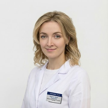
Karpova Maria Andreevna
Experience 15 years
Make an appointment
Karpova
Maria Andreevna
Head of the mammological center
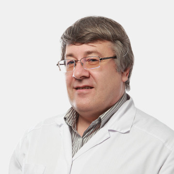
Abalmasov Vladimir Georgievich
Experience 42 years
Make an appointment
Abalmasov
Vladimir Georgievich
Leading specialist in ultrasound diagnostics
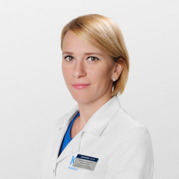
Andreytseva Marina Igorevna
Experience 17 years
Make an appointment
Andreytseva
Marina Igorevna
Ultrasound Diagnostic Doctor
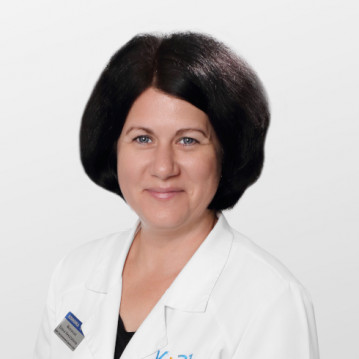
Malakhova Elena Nikolaevna
Experience 16 years
Make an appointment
Malakhova
Elena Nikolaevna
Ultrasound Diagnostic Doctor
Maso Michael Lvovich
Experience 19 years
Make an appointment
Maso
Michael Lvovich
Mammologist, radiologist, ultrasound diagnostician
Mikula Lada Nikolaevna
Experience 32 years
Make an appointment
Mikula
Lada Nikolaevna
Ultrasound Diagnostic Doctor
Shirokih Pavel Nikolaevich
Experience 17 years
Make an appointment
Shirokih
Pavel Nikolaevich
Ultrasound and Prenatal Diagnostic Doctor
Sokolova Alena Victorovna
Experience 16 years
Make an appointment
Sokolova
Alena Victorovna
Ultrasound Diagnostic Doctor
Panfilova Ekaterina Alexandrovna
Experience 21 year
Make an appointment
Panfilova
Ekaterina Alexandrovna
Ultrasound Diagnostic Doctor
Baranova (Vorfolomeeva) Oksana Vladimirovna
Experience 19 years
Make an appointment
Baranova (Vorfolomeeva)
Oksana Vladimirovna
Ultrasound diagnostics doctor
Kozlova Alla Valerievna
Experience 21 year
Make an appointment
Kozlova
Alla Valerievna
Ultrasound diagnostics doctor
Kuznetsova Irina Aleksandrovna
Experience 23 years
Make an appointment
Kuznetsova
Irina Aleksandrovna
Ultrasound Specialist
Shpilevoy Nikolay Yurievich
Experience 20 years
Make an appointment
Shpilevoy
Nikolay Yurievich
Cardiovascular surgeon, surgeon, ultrasound specialist
Gorbik Lyubov Gennadievna
Experience 37 years
Make an appointment
Gorbik
Lyubov Gennadievna
Ultrasound diagnostics doctor, pediatric
Gromovenko Elena Yurievna
Experience 31 year
Make an appointment
Gromovenko
Elena Yurievna
Surgeon, oncologist (mammologist), ultrasound diagnostician, radiologist
Petrovskaya Yana Valerievna
Experience 18 years
Make an appointment
Petrovskaya
Yana Valerievna
Leading ultrasound diagnostic physician
Vozmitel
Daria Andreevna
Ultrasound Diagnostics Doctor
Gumerova Dinara Radikovna
Experience 4 years
Make an appointment
Gumerova
Dinara Radikovna
Obstetrician-gynecologist, ultrasound doctor
Lapaev Igor Vladimirovich
Experience 10 years
Make an appointment
Lapaev
Igor Vladimirovich
Ultrasound diagnostics doctor
Cherepanova Anastasiya Igorevna
Experience 7 years
Make an appointment
Cherepanova
Anastasiya Igorevna
Ultrasound Diagnostics Doctor
Melkonyan Lia Eduardovna
Experience 13 years
Make an appointment
Melkonyan
Lia Eduardovna
Oncologist, oncologist-mammologist, surgeon, radiologist, ultrasound diagnostician
Mebonia Sofia Teymurazovna
Experience 20 years
Make an appointment
Mebonia
Sofia Teymurazovna
Functional Diagnostic Doctor
Belevskaya Anna Andreevna
Experience 13 years
Make an appointment
Belevskaya
Anna Andreevna
Functional Diagnostic Doctor
Achba Maya Otarovna
Experience 18 years
Make an appointment
Achba
Maya Otarovna
Radiologist, ultrasound diagnostician, oncologist-mammologist
All specialists
2GIS Award
2GIS Award

This award is given to clinics with the highest ratings according to user ratings, a large number of requests from this site, and in the absence of critical violations.

«Good place» according to Yandex

This award is given to clinics with the highest ratings according to user ratings. It means that the place is known, loved, and definitely worth visiting.

Our doctors are laureates of the ProDoctors Award

The ProDoctors portal collected 500 thousand reviews, compiled a rating of doctors based on them and awarded the best. We are proud that our doctors are among those awarded.

Make an appointment at a convenient time on the nearest date

Price

Reception
Price
Breast ultrasound
from 7 310 ₽

Appointment to the doctor

Fill out the form, our managers will contact you within 15 minutes

Reviews

It's not the first time I've had an ultrasound at Petrovskaya Yana Valeryevna. Excellent specialist, highly qualified. In addition to professionalism, it should be noted that Yana Valeryevna is a very attentive, friendly and sympathetic doctor. Her reception leaves the best impressions. Thank you very much!
07.07.2026
Mariya K.
I am being observed by Dr. Maso M.L. The doctor is excellent, professional, and empathetic. He is always involved, takes his time and answers all questions in great detail. I also want to mention the work of the mammology department manager, Ksenia. It is pleasant to communicate with Ksenia, she is always friendly to the patient, you can contact her for any reason and know that Ksenia will make every effort to organize everything at the highest level. The only negative aspect relates to the reception on the ground floor. Unfortunately, the long wait and unhurried administrators spoil the impression of the clinic.
07.07.2026
Alesya A.

About doctor:

Maso Michael Lvovich

I liked the attentive and caring attitude, the specialist answered my questions in simple, understandable language.
06.07.2026
Alina G.
The good doctor
01.07.2026
Elena B.

About doctor:

Vozmitel Daria Andreevna

The best ultrasound technician
25.06.2026
Anush D.

About doctor:

Mikula Lada Nikolaevna

A wonderful doctor. She is very polite and very helpful. She told me everything, explained everything, and answered all my questions.
23.06.2026
Oksana S.
I have been seeing Elena Nikolaevna since 2016. Very attentive, competent, friendly and highly qualified specialist! Thank you so much for your work!
23.06.2026
Vladimir F.
Many thanks to the Department of Mammology, laboratory assistants, manager and doctors Maso M.L. and Achbe M.O.
22.06.2026
Marina Sh.
Почему К+31?
К + 31 — full-cycle multidisciplinary medical centers, including the possibility of providing medical services of European quality level.
К + 31 — are leading doctors and diagnostics using high-tech equipment from world manufacturers (Karl Storz, Olympus, Siemens, Toshiba, Bausch&Lomb, Technolas, Zeiss, Topcon).
К + 31 — is ethical. The staff of K+31 clinics maintain open relationships with patients and partners. An individual approach to each patient is the basis of our service standards.
К + 31 — is modernity. On call 24/7: call center operators will answer your questions at any time and book you an appointment with doctors. Contact us by phone, through the feedback form on the website and Max.

Our clinics

Address K+31 on Lobachevskogo

st. Lobachevskogo, 42/4

Contacts

+7 499 999-31-31

Opening hours

Mon-Fri: 08:00 – 21:00
Weekend: 09:00 – 19:00

Address K+31 Petrovskie Vorota

1st Kolobovsky pereulok, 4

Contacts

+7 499 999-31-31

Opening hours

Mon-Fri: 08:00 – 21:00
Sat-Sun: 09:00 – 19:00

Address K+31 West

Orshanskaya, 16/2; Ak. Pavlova, 22

Contacts

+7 499 999-31-31

Opening hours

Mon-Fri: 08:00 – 21:00
Sat-Sun: 09:00 – 18:00
K+31 Petrovskie Vorota
Didn't find the service you were looking for?

Экстренная помощь