
Vacuum aspiration biopsy (VAB)
When a node is found in the mammary gland, it is important for a woman to quickly understand its nature. A vacuum aspiration biopsy gives this answer accurately and carefully. This is a minimally invasive procedure that does not leave a noticeable scar and does not require hospitalization.
The team of mammologists "K+31" conducts it in Moscow under the supervision of ultrasound, MRI or X-ray. Below we explain when a neoplasm biopsy is needed, how the intervention goes and what to expect after it.

specialists

equipment

treatment







For what findings and diagnoses do we recommend a neoplasm biopsy
The doctor always determines the indications after the examination, and the decision is not made based on just one picture. Most often, a neoplasm biopsy is needed in such situations:
- A dense knot that is palpable or visible in the images
- Unclear changes according to mammography and ultrasound
- Growth of previously stable education
- Suspicious microcalcinates
- Category BI-RADS 3-5 based on survey results
- Enlarged lymph nodes of the axillary region
Let's analyze the main findings in more detail so that it is clear why in each case we recommend clarifying the diagnosis by taking tissue.

Fibroadenoma and other benign tumors
We make the decision on removal together with the patient, weighing the size of the node and the complaints.
Suspected breast cancer
If the scans are alarming, you can not delay. If breast cancer is suspected, a biopsy provides material for histology and immunohistochemistry, and therefore the basis for a treatment plan.
The sooner the diagnosis is confirmed or eliminated, the safer both the doctor and the patient feel. At the same time, the biopsy itself does not mean a diagnosis — suspicion is often not confirmed.
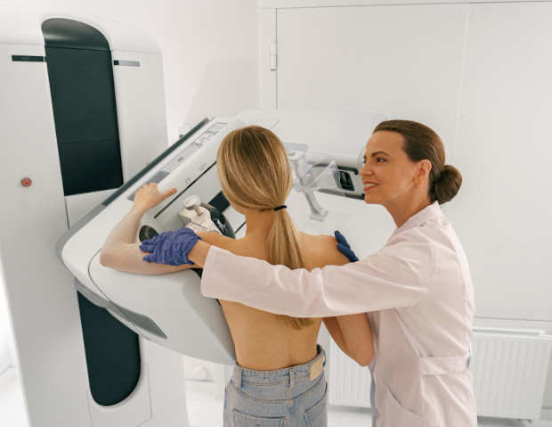
Unclear changes according to mammography and ultrasound
Sometimes education is difficult to assess unambiguously from photographs. Then tissue sampling puts an end to the question, and not repeated blind observations. It gets rid of from months of waiting and worrying too much.
Control and monitoring of previously identified formations
A node that changes from inspection to inspection requires clarity. An imaging-controlled biopsy helps to understand whether it is worth continuing to monitor or whether it is time to act. This way we don't miss important changes.
Lymph nodes of the axillary region
Enlarged axillary lymph nodes are also amenable to targeted tissue sampling. This is important for an accurate assessment of the prevalence. the process, if we are talking about a malignant tumor.
Types of vacuum biopsy in the K+31 clinic
Education should be clearly visible to the doctor during the procedure, otherwise there is no need to talk about accuracy. Therefore, at the K+31 clinic, we We use three navigation methods and choose the one that suits you. Let's briefly compare them in the table:
| Control method | When applied | Features of the procedure |
|---|---|---|
| Under ultrasound navigation | Nodes visible on ultrasound | Fast, comfortable, without radiation load |
| Under MRI control | Changes visible only on tomography | High precision, need contrast enhancement |
| Under X-ray control | Microcalcinates on the mammogram | Stereotactic targeting of small inclusions |
Let's analyze each method in more detail so that it is clear in which situations it helps.
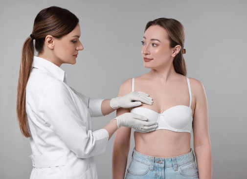
Vacuum aspiration biopsy (VAB) under ultrasound navigation
If the node is visible on ultrasound, we work under ultrasound navigation. It is fast, comfortable and without radiation exposure, and the doctor sees the needle in real time and fully controls every movement. We choose this option most often because it is the most gentle.
Vacuum aspiration biopsy (VAB) under MRI control
Some of the changes are noticeable only on magnetic resonance imaging. In such cases, we perform a biopsy under MRI control — this is the most sensitive navigation that helps to get to hidden areas. Contrast enhancement is usually performed before the procedure.Vacuum aspiration biopsy under X-ray control (stereotactic)
Stereotactic access is used for microcalcifications that are visible only on a mammogram. An X-ray-controlled biopsy precisely points the needle at the smallest inclusions that cannot be felt. The method is especially valuable for early diagnosis when the changes are still very small.
How do we perform a vacuum biopsy
The patient's entire journey is thought out and takes a little time. A breast biopsy under visual control is a few clear steps, not a long operation. We accompany the woman at every stage, from the first interview to the issuance of the report, and answer all questions in advance.
Diagnosis and preparation for intervention
First, the mammologist examines the pictures and examines the breast. Then we select the navigation method and explain the procedure in detail. Special complex training is usually not required, it is enough to take the results of previous examinations with you. If you are taking blood thinning medications, it is important to tell this in advance.
General information
Our advantages in conducting VAB at the K+31 clinic
Vacuum biopsies are performed in many places, but the result depends on the team and equipment. Here's what the patient gets at the K+31 clinic:
- Three navigation methods — ultrasound, MRI and X-ray — in one clinic
- Minimally invasive technique without stitches and noticeable scars
- A team of mammologists, radiologists and morphologists
- Own histological laboratory
- Clear support before receiving the diagnosis
- The opportunity to continue treatment if necessary
This format saves time and relieves anxiety: the patient goes through the entire route in one place, without unnecessary travel and repeated consultations in different centers. A single route is especially appreciated by busy women who value every hour.
Minimally invasive access and aesthetic result
We're working through one small puncture. Minimally invasive biopsy does not require sutures and leaves a barely noticeable mark, which is especially important for a woman. The breast retains its usual shape, and recovery takes place quickly.
Three navigation methods in one clinic
Not every education is equally visible in different studies. The presence of ultrasound, MRI and X-ray under one roof allows you to choose the most accurate control without unnecessary travel and loss of time. This eliminates the need to look for the necessary equipment in other centers and speeds up the diagnosis.
A team of mammologists, radiologists and morphologists
The result of the biopsy is a collaborative effort. An oncomammologist, a radiologist, and a morphologist together provide precision at every stage, from pointing the needle to incarceration under the microscope. We discuss controversial cases together so as not to miss a single detail.


Frequently Asked Questions
We have collected what women ask most often before the procedure.
How does a vacuum biopsy differ from a conventional puncture?
Does it hurt to have breast cancer?
The procedure is performed under local anesthesia. The patient feels only a slight prick during anesthesia, there is no further pain. We use modern anesthetics and monitor the condition at every stage.
Which control method should I choose: ultrasound, MRI or X-ray?
The choice depends on how the education is better seen. Ultrasound node — we work under ultrasound navigation, changes only on tomography are under MRI control, microcalcifications on mammogram are under X—ray control.
How quickly will the histology result be ready?
How safe is a vacuum biopsy?
This is a minimally invasive procedure with minimal risk of complications. The puncture is only two to three millimeters, stitches are not needed, and the risk of hematoma or infection is extremely low.
Our doctors









This award is given to clinics with the highest ratings according to user ratings, a large number of requests from this site, and in the absence of critical violations.

This award is given to clinics with the highest ratings according to user ratings. It means that the place is known, loved, and definitely worth visiting.

The ProDoctors portal collected 500 thousand reviews, compiled a rating of doctors based on them and awarded the best. We are proud that our doctors are among those awarded.
Make an appointment at a convenient time on the nearest date
Price

Appointment to the doctor
Reviews




Our clinics


Application “Personal Account K+31”












What is a vacuum aspiration biopsy and when is it needed?
This is a way to take tissue through one small puncture. A special vacuum-assisted system takes several whole tissue columns, rather than individual cells, as with a conventional puncture. This material allows you to conduct a full-fledged histological examination and make an accurate diagnosis. We offer a VAB when there are not enough examinations and photographs, and the issue of the nature of education needs to be finally closed.
The method solves two problems at once. It confirms or excludes the malignant process and, with small benign nodules, often removes them completely. Therefore, a breast biopsy in this way is both diagnostic and, in many cases, therapeutic. For a woman, this means fewer interventions and less anxiety.