Dental granuloma: symptoms, diagnosis and treatment
A dental granuloma is an inflammation of the root apex. Despite its small size—usually 5–8 mm—it is a constant source of infection in the body. Treating a dental granuloma offers the opportunity to save the tooth and stop the spread of infection.

specialists

equipment

treatment


Causes of dental granuloma

The main factor in the development of pathology is infection. A healthy tooth is reliably protected by enamel, but as soon as the integrity of the hard tissues is compromised, bacteria gain access to the deep structures.
Advanced caries, pulpitis, periodontitis
Causes of dental granuloma include caries and nerve destruction. If the pain is tolerated, pus will drain into the root canals.
Root canal infection
Granulomas can also appear in previously treated teeth. Anaerobic microbes multiply in the remaining cavities. Gradually, their activity leads to the development of chronic inflammation at the root apex.
Tooth trauma and other provoking factors
Mechanical damage is also a possible cause. A blow, a fall, an improperly fitted crown, or the habit of chewing on hard objects can cause a rupture of the neurovascular bundle. This creates an ideal environment for bacteria.

Symptoms of a tooth granuloma
A granuloma doesn't hurt for a long time. You can live for years without realizing anything is wrong with your tooth. But as soon as it grows in size, a flare-up occurs.
Early stage signs
In the initial stages, symptoms of a tooth granuloma are virtually absent. Sometimes, a slight discoloration of the crown (it becomes grayish) or occasional discomfort when eating hot foods may be observed.
Symptoms during an exacerbation of inflammation
- Pain when biting
- Redness and swelling of the gums
- Feeling that a tooth has become higher than the others
- Bad breath
When to urgently see a doctor
Self-medication, the use of heating pads, or rinsing with alcohol-based tinctures only accelerate the spread of pus into the soft tissues.
You should see a dentist if you experience:
- Pain that is not relieved by analgesics
- Cheek swelling (flux)
- Fistula with pus
- Body temperature above 37.5°C
- Difficulty opening the mouth
If swelling and purulent discharge increase, immediate treatment of the infection in a dental clinic is required.
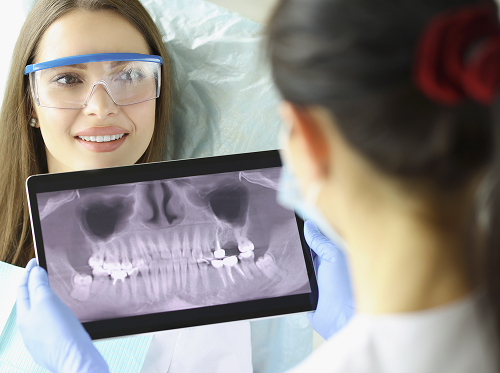
Diagnosis of dental granuloma
Since granulomas are not visible visually, diagnosis requires a comprehensive approach and the use of imaging techniques.
Examination and collection of complaints
During the examination, the dentist checks for the presence of cavities, old fillings, or crowns. Percussion (tapping the tooth) is mandatory; in the presence of a granuloma, this procedure often causes a painful reaction. The condition of the mucous membrane is also assessed for the presence of sinus tracts or infiltrates.
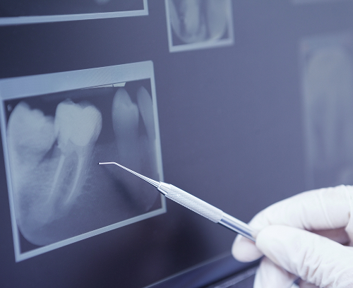
X-ray and CBCT
The gold standard for detecting hidden inflammation is radiographic diagnostics.
- A dental X-ray (a targeted image) reveals a darkening at the root apex with clear boundaries. However, a flat image does not always provide complete information about the shape and extent of the lesion.
- Dental CBCT (computed tomography) is the most accurate method. Three-dimensional reconstruction allows for a full view of the tooth, assessing the number of canals, their patency, and the true size of the inflammatory lesion in the bone. Diagnosing dental granulomas with CBCT eliminates errors in treatment planning.
How to distinguish a granuloma from a cyst and other inflammatory lesions?
A granuloma is up to 0.5 cm in diameter and has distinct borders on the image. A cystogranuloma reaches 0.8–1 cm. Anything larger than 1 cm is considered a cyst.
General information
Dental granuloma treatment methods
| Method | When to use | Advantages | Limitations | What's important after treatment |
|---|---|---|---|---|
| Endodontic treatment of dental granuloma | Primary inflammation, patent root canals | Tooth preservation, low trauma | Requires time and several visits | Follow-up X-rays after 6 months |
| Retreatment of root canals | Recurrence under an old filling, poor-quality obturation | Chance to save the tooth, elimination hidden infection | High complexity, risk of perforation of the walls | Regular hygiene, monitoring by a doctor |
| Root apex resection | Large lesion, inability to pass the canal | Removal of infection without tooth extraction | Surgery, presence of sutures | Postoperative care, antiseptics |
| Tooth extraction | Complete destruction of the root, vertical crack | Guaranteed elimination of the source of infection | Tooth loss, risk of displacement of adjacent units | Implantation or prosthetics |
Conservative treatment through root canals
This is the most common method. The dentist's goal is to completely sterilize the internal space of the tooth. Through the created access, the dentist removes tissue decay and bacterial waste products. A special medicinal paste based on calcium hydroxide is placed in the canals, which has a powerful antiseptic effect and promotes bone regeneration.
Retreatment of a previously treated tooth
If inflammation has developed under an old filling, root canal retreatment is performed. The dentist removes the filling, old posts or fragments, and then performs repeated disinfection. Only after complete cleaning can the inflammatory focus be removed.
Root apex resection
In cases where the canal is blocked (for example, by a broken instrument or very hard cement), and a granuloma remains at the root, a surgical procedure is used. Root apex resection involves removing only the infected portion of the root and the granuloma itself through a small incision in the gum. This allows the tooth to be preserved as a functional unit.
When is tooth extraction really necessary?
Unfortunately, treatment is not advisable in all situations. Extraction is recommended if:
- The tooth is decayed deep beneath the gum and cannot be restored with a crown.
- A vertical root crack is detected.
- The granuloma is so large that it has destroyed most of the tooth's bone support.
- The canals are completely obstructed, and surgical access is impossible.


Prevention means:
- Visiting the dentist every six months
- Treating cavities at the first sign of tooth decay
- Brushing your teeth twice a day, professional cleaning
- X-rays of teeth with old fillings
Frequently asked questions
Can a granuloma be cured with antibiotics?
Is it necessary to extract a tooth with a granuloma?
Can a granuloma go away on its own?
How long does granuloma treatment last?
Our doctors


This award is given to clinics with the highest ratings according to user ratings, a large number of requests from this site, and in the absence of critical violations.

This award is given to clinics with the highest ratings according to user ratings. It means that the place is known, loved, and definitely worth visiting.

The ProDoctors portal collected 500 thousand reviews, compiled a rating of doctors based on them and awarded the best. We are proud that our doctors are among those awarded.
Make an appointment at a convenient time on the nearest date
Price

Appointment to the doctor
Reviews


Our clinics

Application “Personal Account K+31”


What is a dental granuloma?
A granuloma is not a tumor, but rather a protective response of the body. When an infection spreads beyond the root, the immune system attempts to contain the invader by constructing a connective tissue barrier.
Where is a granuloma located and how does it form?
A granuloma is located at the root of the tooth, specifically at the apex (apex). The process of formation begins with the penetration of microbes from the infected pulp through a narrow opening into the root and then into the surrounding periodontium. In response to constant irritation from bacterial toxins, connective tissue cells begin to actively divide. Gradually, a dense nodule forms at the site of the destroyed bone. While the immune system restrains bacterial growth, the process is chronic, but any weakening of the body's defenses (colds, stress, hypothermia) can lead to an acute phase of the granuloma.
What is the difference between a granuloma and a cyst?
There are three stages of inflammation: granuloma, cystogranuloma, and cyst. The main difference is size and structure:
A granuloma is easier to treat. A cyst requires surgery.