Treatment of Dental Enamel Hypoplasia
Dental enamel hypoplasia is a non-carious tissue lesion that occurs as a result of a disruption in the process of tooth formation. Unlike dental caries, which destroys the tooth after it has erupted, this pathology develops during the mineralization and formation of the protein matrix. In this condition, enamel may be thinned, discolored, or completely absent in certain areas. Timely treatment of enamel hypoplasia not only restores the aesthetics of a smile but also protects the deep tissues of the tooth (dentin and pulp) from bacteria and temperature irritants.

specialists

equipment

treatment


How does enamel hypoplasia manifest itself?
Manifestations of the disease depend on the stage and form of the disorder. Dentists classify defects based on visual signs and the patient's subjective sensations.
External Signs
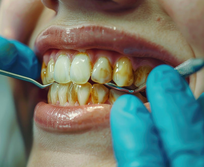
Visually, the pathology manifests as a change in the structure or color of the tooth surface. The following may be observed on the enamel:
- White spots or yellowish areas with a smooth, shiny surface
- Horizontal grooves on the teeth, encircling the crown
- Pinpoint depressions (pits), which may become pigmented and brown over time
- General thinning of the enamel, causing yellowish dentin to show through.
Patient Complaints and Sensations
Tooth sensitivity appears first. It manifests as a sharp, short-term pain upon contact with cold, hot, sour, or sweet foods. In advanced cases, when the enamel layer is minimal, pain can even occur when inhaling cool air.
Causes of enamel hypoplasia

It's all about the ameloblast cells that "build" enamel. If a malfunction occurs during their function, the cells stop secreting the necessary material. As a result, the tooth's protective layer either doesn't form at all or is too thin and uneven.
Causes in Children During Tooth Formation
Systemic causes of enamel hypoplasia are rooted in the health of both mother and child during critical periods of development. These include:
- Severe toxicosis, maternal infectious diseases during pregnancy (rubella, acute respiratory viral infections)
- Rhesus incompatibility
- Birth injury or severe prematurity
- Calcium and phosphorus metabolism disorders in the first years of a child's life (rickets)
- Acute childhood infections and endocrine disorders
Causes of localized damage to individual teeth
In some cases, not the entire dentition is affected, but a specific tooth. This occurs due to mechanical trauma to the bud of a permanent tooth or as a result of chronic inflammation of a primary tooth (periodontitis), the infection from which spreads to the deep tissues of the jaw. As a result, the permanent tooth erupts with a defect.
Types of enamel hypoplasia
Dentists distinguish three main categories of the disease depending on the prevalence of the process.
Systemic Hypoplasia
Systemic enamel hypoplasia is characterized by damage to a group of teeth that formed during the same period. If the disruption was short-lived, the defect appears as a horizontal line. If the disease is long-lasting, large areas of the crowns are affected. Symmetrical teeth (for example, both central incisors) are usually affected.
Local Hypoplasia
Local enamel hypoplasia affects one or two teeth. Typically, these are premolars. The cause is always a local factor in the jaw tissues, rather than a general health issue. Enamel may become stained or change texture.
Enamel aplasia
Enamel aplasia (complete absence of enamel) is the most severe form of developmental defect. Dentin is left completely unprotected, leading to the immediate development of caries and severe pain.
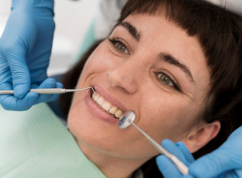
Diagnosing Enamel Hypoplasia
A dentist should make a diagnosis immediately. Each pathology has its own treatment strategy.
Examination and History
Diagnosing enamel hypoplasia primarily involves clarifying the symptoms. The doctor will determine the mother's pregnancy history and ask about childhood illnesses and injuries. During the examination, the dentist will evaluate the symmetry of the lesions, the density of the defect bases, and their reaction to staining.
Differential diagnosis with caries, fluorosis, and enamel erosion
A comparison method is used:
- Caries: single spots, rough tooth surface, stained with methylene blue
- Fluorosis: in this case, there are multiple spots, affecting all teeth, not just symmetrical groups
- Erosion: more common in adults, has a cup-shaped form, and is associated with chemical or mechanical impact on an already erupted tooth
General information
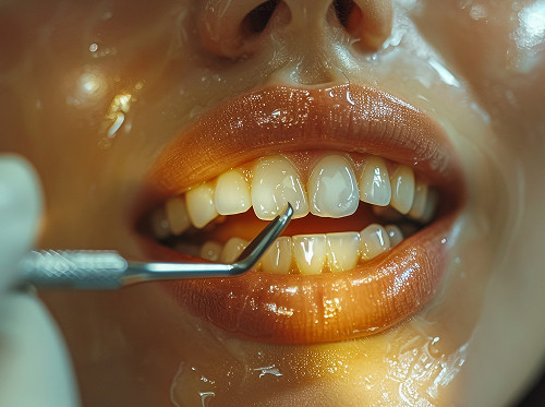
Enamel Hypoplasia Treatment Methods
There are several treatment methods. Let's look at each in more detail.
Remineralizing Therapy
In the initial stages, enamel remineralization is performed. Calcium, phosphorus, and fluoride preparations are applied to the teeth. This will not completely remove the stain, but it will strengthen the tissue structure, reduce sensitivity, and prevent the development of caries.
Aesthetic Correction of Surface Defects
Microabrasion, which involves grinding away a thin layer of the affected enamel and then polishing, can be used to remove white spots.
Composite Restoration
Teeth restoration is performed if there are obvious defects. The dentist fills the cavities with a material similar in color and strength to enamel.
Orthopedic restoration for severe defects
If the defects are too severe, the dentist may recommend installing:
- Veneers: thin ceramic shells for the front teeth that completely conceal defects.
- Crowns: in the case of extensive decay, they protect the tooth from all sides, restoring its function and appearance.


Frequently Asked Questions
What is enamel hypoplasia?
Can enamel hypoplasia be completely restored?
How does enamel hypoplasia differ from caries?
Is enamel hypoplasia dangerous in children?
Our doctors


This award is given to clinics with the highest ratings according to user ratings, a large number of requests from this site, and in the absence of critical violations.

This award is given to clinics with the highest ratings according to user ratings. It means that the place is known, loved, and definitely worth visiting.

The ProDoctors portal collected 500 thousand reviews, compiled a rating of doctors based on them and awarded the best. We are proud that our doctors are among those awarded.
Make an appointment at a convenient time on the nearest date
Price

Appointment to the doctor
Reviews


Our clinics

Application “Personal Account K+31”


Degrees and forms of damage
The clinical picture allows us to classify the disease based on morphological features.
Spotted form
The mildest degree. Clear spots on the enamel are visible on the teeth, usually white or chalky yellow. The surface of the spot remains smooth and dense; the enamel is not mechanically damaged, but its optical properties change.
Sulcate and pitted form
These are more pronounced defects. Pinpoint pits or wavy depressions are visible on the anterior surface. If the grooves are deep, the tooth takes on a specific shape (for example, Hutchinson teeth with a crescent-shaped notch on the incisal edge).
Thinning or absence of enamel
With this form, teeth may appear smaller, have an unnaturally yellow color, and become cone-shaped. Severe sensitivity develops, and chewing becomes difficult due to the fragility of the tissue.