Sialoadenitis Treatment in Moscow
Sialoadenitis is an inflammation of the salivary glands. The patient experiences swelling and general malaise. The inflammation is caused by viruses, tooth decay, and immune system problems.
Timely treatment of sialoadenitis in Moscow allows for rapid pain relief and prevents the disease from becoming chronic or developing into a purulent lesion.

specialists

equipment

treatment


Sialoadenitis refers to damage to the major or minor salivary glands, whether caused by an infectious or non-infectious cause. The salivary gland produces a secretion that is essential for the initial breakdown of food and the protection of the oral mucosa. When its tissues become inflamed, saliva flow is impaired.
Which salivary glands are most commonly inflamed?
Parotid sialoadenitis (mumps) is the most commonly diagnosed type. In this case, swelling is localized in front of and below the auricle. Submandibular sialoadenitis is the second most common type of sialorrhea: pain typically occurs in the lower jaw. Inflammation of the sublingual glands is rare.
How does acute sialoadenitis differ from chronic sialorrhea?
Acute sialoadenitis develops suddenly. The patient develops pain in the salivary gland, fever, and swelling.
With proper treatment, the symptoms resolve without a trace. Chronic sialadenitis is less severe but long-lasting. Periods of remission alternate with exacerbations, during which the gland gradually hardens and its function is irreversibly impaired.
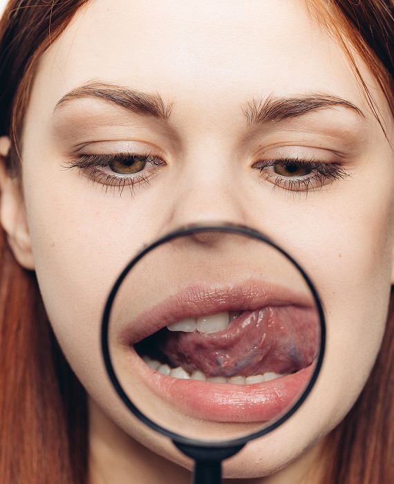
Symptoms of sialadenitis

The clinical presentation of the disease depends on the type of inflammation, but there are a number of common signs that can help suspect the pathology at an early stage.
Early signs: pain, swelling, dry mouth
The first symptom is a feeling of fullness and discomfort. Gradually, swelling of the salivary gland develops, which can alter the contours of the face. Due to impaired secretion, severe dry mouth occurs, and the saliva itself may become viscous or contain flakes.
Symptoms that worsen during eating
A characteristic sign of the disease, especially if there is an obstruction in the duct, is "salivary colic." This is a sharp, stabbing pain when chewing or even at the sight of food. Since eating stimulates saliva production, and an inflamed or clogged salivary gland duct cannot pass it, intra-tissue pressure increases sharply.
Signs of purulent inflammation and complications
If the process progresses, purulent sialadenitis may develop. In this case, the skin over the gland becomes red and hot. Pus may leak from the duct into the oral cavity, causing an unpleasant salty or bitter taste. The patient's condition worsens: fever reaches high levels, and difficulty opening the mouth becomes apparent.
Causes and risk factors

Inflammation rarely occurs on its own; it is most often the result of infection or impaired fluid drainage.
Bacterial and viral infection
Viral sialoadenitis is often caused by influenza viruses, cytomegalovirus, or mumps. Bacterial inflammation is caused by staphylococcus and streptococcus bacteria, often accompanied by poor hygiene or inadequate dental treatment.
Obstruction of the salivary gland and stones
A salivary stone (sialolithiasis) is a common cause of inflammation in the submandibular region. The hard mass blocks the lumen of the canal, causing secretion stagnation. Obstruction of the duct can also occur due to thickening of saliva or a foreign body, which creates an ideal environment for bacterial growth.
Dehydration, decreased salivary flow, concomitant diseases
General dehydration leads to saliva becoming excessively thick. Post-operative patients and those taking diuretics and antihistamines for a long time are most often affected. A weakened immune system and diabetes also increase the risk of salivary gland inflammation.

When to see a doctor
You should see a doctor immediately if you have:
- Severe swelling near the ear or under the jaw
- Pain that gets worse when chewing
- High fever
- Dry mouth and decreased salivation
- Unpleasant taste or discharge from the duct
- Repeated episodes of inflammation
When to seek emergency care
If swelling extends to the neck or lower eyelid, fever If the temperature rises above 38.5°C and the pain becomes unbearable, immediate assistance is required. Severe pain combined with the inability to swallow even water indicates the development of an abscess. In this situation, an oral and maxillofacial surgeon should perform an emergency examination.
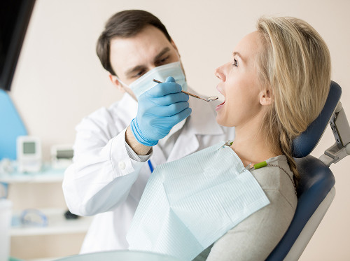
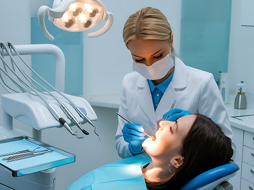
Diagnosis of sialadenitis
Examination, palpation, and collection of complaints and anamnesis
The doctor evaluates facial symmetry, skin color, and the condition of the oral duct openings. Palpation determines the gland's density and the presence of discharge. The dentist determines when the pain began and whether it is related to food intake.
Salivary gland ultrasound and laboratory tests
Salivary gland ultrasound is the "gold standard" for primary diagnostics. This method allows us to visualize the gland's size, the presence of stones, duct dilation, and the presence of purulent cavities. Laboratory blood tests reveal the severity of the inflammatory response in the body.
CT and other methods as indicated
In complex or questionable cases, CT (computed tomography) is prescribed. This examination helps to study the tissue structure in detail and detect even small radiopaque stones. In chronic cases, sialography (X-ray imaging with the injection of a contrast agent into the ducts) may be used.
| Form | What most often provokes | Main symptoms of sialadenitis | What helps clarify the diagnosis | Treatment approach |
|---|---|---|---|---|
| Acute bacterial | Oral infection | High fever, severe swelling, pain | Examination, ultrasound, blood tests | Antibiotics, detoxification |
| Chronic | Duct structure, systemic diseases | Periodic swelling, dryness | Ultrasound, sialography, CT scan | Relapse prevention, saliva stimulation |
| Calculous | Salivary gland stone | Pain with food, dense swelling | Ultrasound, CT scan, duct palpation | Stone removal, diet |
| Suppurative sialadenitis | Advanced acute form | Pus from the duct, skin redness, fever | Ultrasound (search for abscess), examination | Surgical incision, antibiotics |
General information
Sialoadenitis Treatment
Complex treatment of salivary gland inflammation is aimed at eliminating the infectious agent, restoring normal saliva flow, and relieving pain.
Conservative Treatment
In the early stages, anti-inflammatory medications and a salivary-promoting diet (consuming acidic foods, such as lemon, to stimulate secretion) are used. The doctor prescribes antibacterial medications as indicated. It is important to remember that uncontrolled use of antibiotics is prohibited; they must be prescribed by a specialist.
Restoring Salivary Flow in Case of Duct Obstruction
If a duct obstruction is detected, the doctor may perform bougienage—widening the duct with special thin probes. This helps restore normal saliva flow.
Tactics for Salivary Gland Stones
A small salivary gland stone may pass on its own. Large formations require removal. Moscow clinics use minimally invasive techniques, such as sialendoscopy—removal of stones through the duct without skin incisions.
What to do with a purulent abscess
If an abscess is diagnosed, surgery is necessary. The surgeon incise and drains the purulent lesion. After surgery, a course of antibiotics and dressings are prescribed.


Frequently Asked Questions
This material is for informational purposes only. If signs of inflammation appear, an in-person consultation with a doctor is necessary. In Moscow, diagnosis and treatment of sialadenitis are carried out according to current standards.
Can I warm an inflamed gland with a blue lamp or salt?
Which doctor should I see first?
Can sialadenitis resolve on its own?
Can this disease be transmitted to other people?
Our doctors


This award is given to clinics with the highest ratings according to user ratings, a large number of requests from this site, and in the absence of critical violations.

This award is given to clinics with the highest ratings according to user ratings. It means that the place is known, loved, and definitely worth visiting.

The ProDoctors portal collected 500 thousand reviews, compiled a rating of doctors based on them and awarded the best. We are proud that our doctors are among those awarded.
Make an appointment at a convenient time on the nearest date
Price

Appointment to the doctor
Reviews


Our clinics

Application “Personal Account K+31”


Types of sialadenitis
Doctors classify the disease by location and course, which directly influences how sialadenitis is treated in a particular case.
Parotid sialadenitis
Parotid gland involvement is often accompanied by pain radiating to the ear or temple. Swelling can be so severe that the earlobe is raised. It is important to differentiate this condition from lymphadenitis or temporomandibular joint problems.
Submandibular sialadenitis
With this form, the swelling is localized under the soft tissues of the floor of the mouth. A hard, painful mass is often palpated. If the cause is calculous sialadenitis, the stone can be felt with the fingers during self-examination.
Chronic recurrent sialadenitis
This form is characterized by recurring episodes of inflammation several times a year. Between flare-ups, the gland may remain slightly enlarged and dense. Chronic sialadenitis requires a thorough investigation of the cause, as each new relapse replaces the functioning gland tissue with scar tissue.