Mammalogy
Our department of mammology is unique in that it employs multimodal specialists - doctors who can carry out any diagnosis, if necessary, make a biopsy, make a diagnosis and prescribe treatment. And besides, our doctors are excellent psychologists.
They are always ready to listen, encourage, give advice. I want to tell all women that there is no need to be afraid of a mammologist! Moreover, it is important to talk about the importance of regular examinations to your surroundings - mothers, sisters, grandmothers. Modern examinations, both ultrasound and mammography are absolutely painless. Also, everything is healing. Some diseases are more difficult, some are easier, but nonetheless.

To form your own opinion about the quality of services and the atmosphere in the clinic, watch the video about the department of mammology
The Mammology department employs experienced and qualified specialists with positive feedback from patients. You can receive paid medical care, undergo timely diagnosis, get advice and learn the latest news from the field of medicine.
Price
Our doctors


This award is given to clinics with the highest ratings according to user ratings, a large number of requests from this site, and in the absence of critical violations.

This award is given to clinics with the highest ratings according to user ratings. It means that the place is known, loved, and definitely worth visiting.

The ProDoctors portal collected 500 thousand reviews, compiled a rating of doctors based on them and awarded the best. We are proud that our doctors are among those awarded.
Make an appointment at a convenient time on the nearest date
Treatment of diseases
Methods of treatment in the clinic
In mammology, there are different ways to treat breast diseases, which can be divided into 2 categories - surgical and non-surgical. Surgical intervention involves the removal of various types of neoplasms with partial or complete resection of the glandular tissue. It all depends on the size of the tumor, cyst or induration, the exact diagnosis, or the presence of metastases in the case of cancer.
Non-surgical treatment includes different areas:
- Conservative methods consist of drug therapy using vitamins, hormonal or anti-inflammatory drugs. Such treatment is always selected by a doctor, the results depend on the nature of the course of the disease and the individual characteristics of the organism;
- chemotherapy required when breast cancer is detected;
- radiotherapy, used as an additional treatment after breast-sparing surgery and removal of part of the breast. Depending on the type of cancer, the optimal type of radiation therapy is selected to destroy the remaining cancer cells in order to prevent the recurrence of the disease. Also, this type of treatment can be used solo. It all depends on the type of cancer and additional individual factors.
Often in mammology, in the treatment of diseases, an integrated approach is used using different ways to solve a patient's problem. This greatly increases the chances of a complete cure for most diseases. The tactics and treatment plan are selected individually by a mammologist.
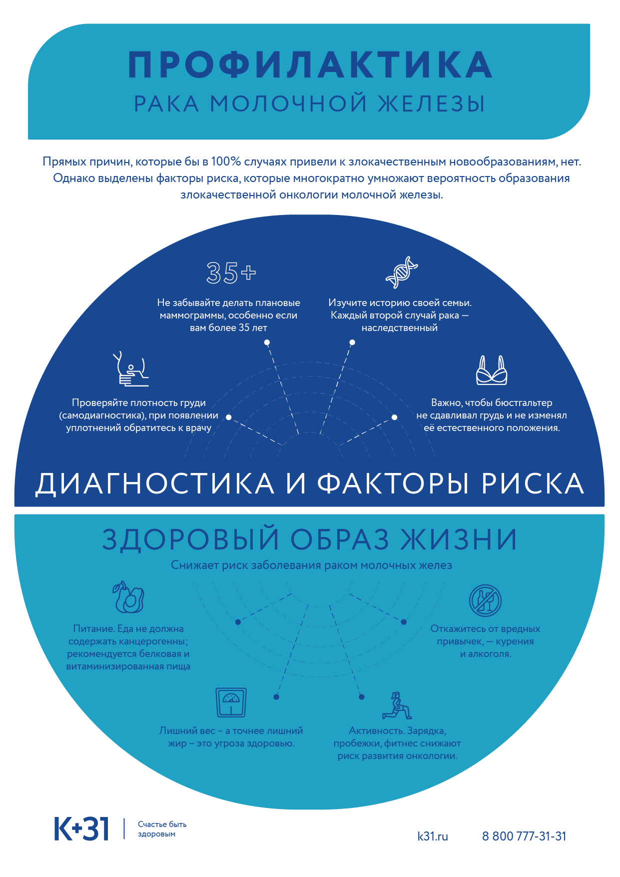
Prevention of breast diseases
Preventive measures of breast diseases are one of the main tools in maintaining health. Among them are:
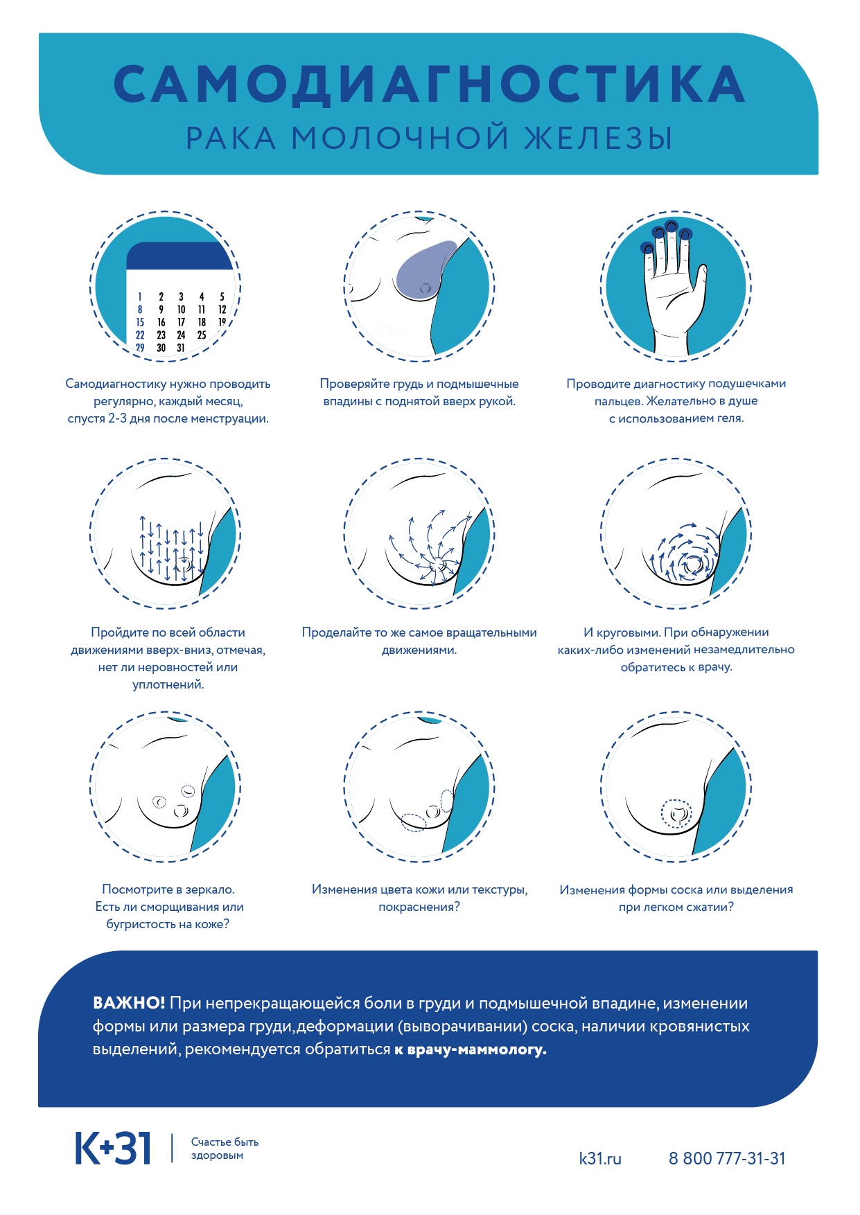
- self-examination of the glands. Regular palpation allows you to detect various seals early, notice uncharacteristic pain sensations and, accordingly, consult a doctor in a timely manner;
- annual ultrasound or mammography (if the patient is over 35);
- control of hormonal levels;
- supporting a proper and healthy lifestyle with a balanced and complete diet;
- Refuse to take hormonal drugs not prescribed by a doctor (including contraceptives).
The number of preventive measures can include testing for mutations in the BRCA1, BRCA2 genes. If a defect is found in them, this significantly increases the risk of developing breast cancer. In these patients, a mastectomy is recommended. Thanks to the possibilities of plastic surgery, instead of glandular tissue, implants can be placed that completely imitate the natural breast. This allows you to maintain aesthetics and prevent the development of breast cancer.
Helpful information
What diseases are treated in the mammology department
K+31 Medical Center in Moscow deals with the diagnosis, treatment and prevention of breast diseases. Our specialists also advise on breastfeeding and help improve milk production. Using modern diagnostic methods, we identify and treat the following diseases:
- Breast cyst and benign tumor
- Mastopathy
- Fibroadenoma
- Breast hypertrophy
- Inflammation of the mammary gland (mastitis)
- Lactostasis
- Pain, congestion in the chest
- Breast cancer
If any diseases are detected, the patient may be referred to the department of oncology, gynecology, phlebology, endocrinology, or urology. Our center has doctors of all specialties.
Diagnostics
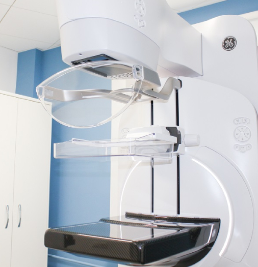
Mammography
Mammography is a standardized procedure consisting of 4 images called projections, two projections of each breast: cranio-caudal or direct and medio-lateral or oblique projections Mammography is performed on specialized x-ray equipment. For the study, a specific technique is used that requires compression of the mammary gland for 5-10 seconds in order to reduce the radiation load on the mammary gland, as well as to obtain high-quality images.
Tomosynthesis
This examination method is also performed in specialized rooms. During the study, standard projections identical to digital mammography are used. The most important difference is the moving X-ray tube, which rotates around the breast and takes several images, which are then reconstructed into sections in 1mm increments. Tomosynthesis can be performed as an independent study, or in combination with standard mammography. In the process of performing a standard mammography, the radiologist decides whether it is necessary to perform an additional study in the tomosynthesis or ultrasound mode. This decision is based on the individual breast x-ray structure, the possibility of using different methods of examination, as well as the characteristics of the changes found during standard mammography.
Contrast mammography
This is a mammogram using iodinated contrast agent. The contrast agent helps to find new blood vessels that appear with the development of a malignant tumor. Your doctor may order a digital contrast mammogram for a variety of reasons, including those listed below.
If you need to be screened for breast cancer. CESM may be particularly helpful in women at increased risk for breast cancer and in women with dense breast tissue.
If it is necessary to evaluate any nodules found in the mammary glands during a medical examination. Contrast digital mammography can help detect breast cancer that cannot be seen on conventional mammograms, especially in women with dense breast tissue.
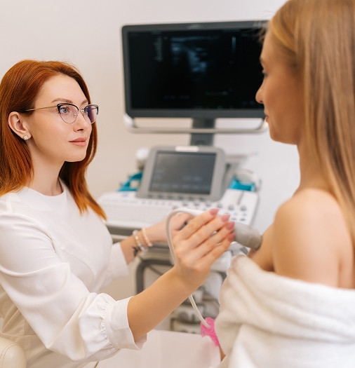
Ultrasound of the mammary glands
It is an indispensable tool in the diagnosis of breast diseases and provides the doctor with information additional to mammography. The addition of ultrasound to mammography improves the radiologist's ability to distinguish benign lesions from those suspicious of breast cancer, thus reducing the number of unnecessary biopsies, especially in dense breast radiographs. Also, ultrasound is a method of primary diagnosis in pregnant and lactating patients.
Breast MRI
MRI or magnetic resonance imaging of the mammary glands with dynamic contrast enhancement is a highly sensitive method that has shown maximum information in the following cases:
- Preoperative planning of the volume of surgical treatment for breast cancer.
- Exclusion of additional foci of breast cancer in the opposite gland.
- Difficult diagnostic cases where there are discrepancies in the results of mammography, ultrasound and clinical examination.
- Control of chemotherapy treatment.
- Implant integrity assessment.
- Diagnosis of postoperative changes in the breast.
- Annual screening for patients at high risk of developing breast cancer.
Biopsy
If necessary, invasive examination methods are used: fine-needle aspiration biopsy, core-needle biopsy, vacuum aspiration biopsy, ductography. A puncture is taken to study the cellular content of a tumor or cyst.
Such studies are carried out under the control of ultrasound, X-ray or MRI and are used to determine the morphology of the tumor and select individual therapy for the patient. To study the tumor in more detail, a blood test (oncogenetics) is used to determine the carriage of the mutated genes BRCA1, BRCA2, CHEK 2, PTEN, p53, CDH-1.
Treatment of breast diseases
Today, almost any breast cancer detected at an early stage can be treated. The tactics of treating a patient is determined by a specialist individually. The K + 31 Clinic provides comprehensive treatment for breast cancer, including surgery and chemotherapy.
Surgery
The volume of the operation directly depends on the size of the formation, the degree of malignancy and the presence or absence of metastases.
Chemotherapy
Medical treatments are also widely used, which include chemotherapy and hormone therapy.
We provide chemotherapy according to the recommendations of NCCN, ASCO, ESMO, using "original" drugs. Therefore, patients tolerate the treatment comfortably.
Radiation therapy
In breast-sparing surgery, where only part of the breast is removed, radiation therapy is performed after the operation, the purpose of which is to destroy additional cancer cells in the remaining breast tissue.
When to contact a mammologist?
A mammologist is a doctor to whom it is important and necessary to visit regularly for examinations.
If there is a family history of breast cancer, mammograms should be done once a year from at least 35 years of age.
On average, annual preventive mammography is recommended for women over 40 years of age. Women over 50 should have a mammogram every six months.
Of course, self-examination is an important stage, and if you feel any changes in the mammary glands (seals, formations, discharge from the nipples), you should immediately consult a doctor.
Patient Library
A woman's mammary glands are a paired organ of external secretion, which throughout life is influenced by various processes, from the menstrual cycle, pregnancy, lactation and ending with menopause. All these and other factors lead to anatomical and physiological changes that can either disappear on their own or lead to the development of pathologies and diseases. All diseases affecting the mammary glands can be divided into several main groups.
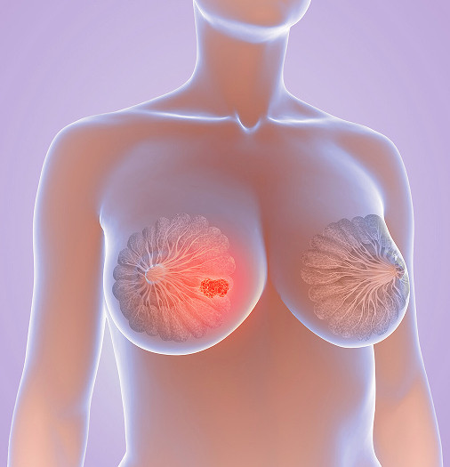
Oncomammology
Oncomammology is a branch of medicine dedicated to the early detection, treatment and prevention of malignant tumors in the mammary glands. According to statistics, among the most common cancers in women, breast cancer ranks first. It is extremely important for girls and women of different ages to undergo preventive examinations in a timely manner in order to detect cancer at an early stage if it occurs.
Pain
Chest pain in women occurs for a variety of reasons, including:
- Premenstrual syndrome
- Burning pain is typical in the first and last trimester of pregnancy
- As a result of cracked nipples associated with inflammatory processes or when feeding a child
- In the presence of benign or malignant neoplasms.
FAQ
What is Better for Early Diagnosis?
When to start the survey?
If nothing bothers you, the examination must be taken from the age of 40. One of the largest international studies showed that performing mammography once a year from the age of 40 reduces mortality from breast cancer by 50%.
If you have changes in your breasts (skin color, shape, nipple discharge, pain).
If there is a genetic predisposition (mutations in the BRCA1 or BRCA2, p53, PTEN, CDH-1, CHEK2 genes have been identified), mammography must be performed starting at the age of 35 in addition to the annual magnetic resonance imaging.
What symptoms to look for?
If you notice:
- pain in the mammary glands not associated with the menstrual cycle;
- redness of the skin, changes in the shape of the mammary gland or nipple;
- discharge from the nipples, not associated with the birth of a child;
- formation that can be felt in the chest.
You need to see a doctor and have a mammogram.
Am I at risk for developing breast cancer?
The main risk factors for developing breast cancer are gender and age. It is for this reason that starting from the age of 40 it is recommended to undergo an examination annually.
The risk also increases if:
- There is a genetic predisposition (the presence of mutations in the genes BRCA1 and BRCA2, p53, PTEN, CHEK-2, CDH-1, which increase the risk of developing breast cancer). Determined by genetic analysis.
- You have been treated for breast cancer in the past.
- Your mom, grandmother, sister or daughter has been diagnosed with breast, ovarian, pancreas, melanoma cancer.
- You had radiation therapy to the chest area at the age of 10-30.
What if I'm at risk but under 40?
If you are at high risk of developing breast cancer, you should start the examination at the age of 25. Annual contrast-enhanced breast MRI is the recommended method. From the age of 35, annual digital mammography is recommended in addition to breast MRI.
Will mammography hurt?
At the time of fixation of the mammary gland with a plate, slight discomfort is possible. To reduce discomfort, you need to relax as much as possible and follow the instructions of the radiologist.
If you were uncomfortable with a previous study, try scheduling your next study on days 7-12 of your menstrual cycle.
And remember: the more compression, the lower the radiation dose and the higher the image quality.
Why check up regularly?
The main purpose of the annual examination is to detect breast cancer at stages 0 and I.
According to the World Health Organization, every 8 women in the world develop breast cancer. However, at an early stage (0 and I) 99% of women live without recurrence of the disease, 98% of women undergo organ-preserving surgery, which allows preserving the aesthetic side of life.
Cystic pathologies
Among the frequent pathologies of the mammary glands, cystic formations are distinguished. They are non-tumor in nature, represent the expansion of ducts filled with fluid. They are found during palpation, echography, pneumocystography, etc. They are successfully treated with conservative treatment, with constant monitoring of the dynamics. Surgical intervention is allowed if the doctor has found signs of malignancy of the cyst.
Among cystic pathologies, there are:
- Mammary adenoma. It appears mainly in young patients and is the result of a hormonal imbalance in the body. It looks like a formation of a spherical or spherical shape, quite elastic, located near the surface of the gland. Adenoma can be multiple or solitary, affects one or both breasts, is treated surgically;
- galactocele, differs from ordinary cysts in the almost complete absence of symptoms. It is most often detected when it reaches a large size or during a mammogram, MRI, ductography, or ultrasound. If the galactocele is small, no treatment is required. With an increase in diameter, removal is performed.
Inflammatory pathologies
This large group of diseases affects the mammary glands of women of different ages. The most common option is mastitis, its types:
- non-lactation acute or chronic form. It is the result of bruises, burns or hypothermia;
- lactational, appears with lactostasis during breastfeeding;
- galactophoritic, consists in inflammatory processes in the milk ducts;
- purulent. With it, a phlegmon or abscess is observed, which gradually degenerates into seals in the mammary gland;
- non-purulent, serous sweating of tissues.
For mastitis of any kind, pain of varying severity, local swelling in the tissues, the appearance of seals that lend themselves well to palpation, periodically appearing fever, as well as weakness and signs of poisoning of the body, are characteristic.
Pathologies after injuries, operations or illnesses
The seals and cysts can appear as a result of breast injuries and initially do not pose a health hazard. If the patient did not turn to the clinic for diagnosis and treatment in time, complications may occur:
- fat necrosis, manifested in focal tissue death, accompanied by the appearance of painful seals. These are benign formations cured by conservative methods;
- calcifications represent the accumulation of calcium salts in the gland;
- capsular contractures consist in the growth of fibrous tissue around the implant installed during mammoplasty.
Those who have undergone breast augmentation with polyacrylamide gel may experience nodules from it that move not only in the chest, but also to other parts of the body.

To get a consultation
Branch news

In October, the K+31 clinic network traditionally supports the international Pink October movement, which highlights the importance of prevention and early detection of breast diseases.

This year, the K+31 medical center chain became an official partner of the "Dalshe" charitable foundation. We support the annual "Pink October" campaign, which aims to highlight the importance of early breast cancer detection.


Radiologist and oncologist-mammologist from the mammology center of the K+31 Clinic. Nadezhda Samvelovna was awarded the All-Russian ProDoctors Prize 2023. She took third place as the best radiologist in Moscow.


Our clinics

Application “Personal Account K+31”


Mammology K+31