Cyst in the Nose and Paranasal Sinuses
Benign tumors in the maxillary, frontal, ethmoid, or sphenoid sinuses are often cysts. These formations appear as thin-walled capsules filled with fluid, mucous, or purulent fluid. This is one of the most common types of ENT lesions. Get treatment for paranasal sinus cysts at the K+31 Clinic.

specialists

equipment

treatment


A sinus cyst is a formation with thin and elastic walls, which is filled with fluid from the inside. The size and location of cysts in the sinus may vary. As a result, the manifestations of cysts may vary from patient to patient.
Inside the mucous membrane, humans have special glands that produce mucus throughout a person’s life. The glands have their own excretory ducts that open onto the surface of the mucous membrane. If, due to an inflammatory process (or for another reason), the gland duct closes, then it stops working, but mucus continues to be produced. The mucus does not find a way out, the walls expand under pressure, which leads to the formation of a spherical container. Most often, cysts form in the area of the maxillary sinuses.
Symptoms of a cyst

The most common symptoms of a cyst are:
- Difficulty breathing. The cyst blocks part of the sinus or presses on the nasal passage.
- A feeling of pressure. This occurs in the cheek, forehead, and bridge of the nose, and intensifies when tilting the head.
- Nasal discharge. This may be mucous or purulent if infected.
- Discomfort in the upper jaw, pressure on the eye. A feeling of heaviness when chewing or pain radiating to the teeth.
- Periodic or persistent headaches. These are associated with impaired ventilation and increased pressure in the sinus.
- Mucus dripping down the back of the throat. Causes a sore throat, cough, and a lump in the throat.
Headaches may appear or intensify when diving deep or ascending rapidly. This is due to changes in air pressure in the sinuses.
If the cyst is located in the sphenoid sinus, symptoms characteristic of sphenoiditis may occur. These include pain in the back of the head, double vision, and decreased vision.
Symptoms depend on the location and size of the cyst and the structure of the maxillary sinus. Furthermore, the condition may be asymptomatic. Small cysts do not interfere with breathing or cause pain, so they can remain undetected for a long time.
Diagnosis and treatment of paranasal sinus cysts
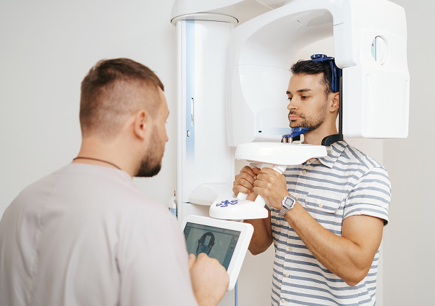
X-ray methods are used for diagnosis. The most optimal is CT (computed tomography) of the nasal cavity and paranasal sinuses, which allows you to determine the size of the cyst and its location with millimeter accuracy.
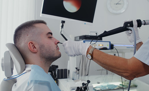
Treatment of cysts in the paranasal sinuses is possible only with the help of surgical methods. Today, the gold standard of surgery is the use of endoscopic techniques in combination with the FESS technique. In the Otorhinolaryngology Department of the K+31 Clinic, endoscopic instruments are used, which allows the doctor to monitor the entire progress of the operation on the monitor in real time and see the microstructure of the nose. The operation is performed through the nose, which helps avoid incisions on the face. The method has virtually no contraindications, rarely has complications, reduces the time of hospital stay (patients are discharged on the same day) and allows you to return to the normal rhythm of life as quickly as possible.
General information about treatment
Symptoms of a radicular cyst
A radicular cyst can exist asymptomatically for years, gradually increasing in size. The patient may notice swelling in the area of the tooth root. Problems arise when the contents of the cyst, for one reason or another, suppurate and a fistulous tract is formed. In the case of inflammatory phenomena, aching pain appears in the tooth, a feeling that the “tooth has grown”; an opening appears outside, in the gum area, from which the contents are released (the opening of the fistulous tract).


Prevention
To prevent sinus cysts, it's important to take preventative measures:
- Treat a runny nose promptly to prevent chronic inflammation of the mucous membrane and sinus blockage.
- Strengthen your immune system. A balanced diet, exercise, physical activity, and adequate sleep help the body fight infections.
- Maintain healthy teeth and gums. Infections of the upper teeth can spread to the maxillary sinuses, leading to the development of cysts.
- Regular examinations allow you to detect pathology before symptoms appear.
To reduce the risk of mucous membrane swelling and sinus blockage, avoid exposure to allergens with allergic rhinitis. Maintaining clean and humid air helps the mucous membrane function normally and reduces the likelihood of inflammation.


Our doctors


This award is given to clinics with the highest ratings according to user ratings, a large number of requests from this site, and in the absence of critical violations.

This award is given to clinics with the highest ratings according to user ratings. It means that the place is known, loved, and definitely worth visiting.

The ProDoctors portal collected 500 thousand reviews, compiled a rating of doctors based on them and awarded the best. We are proud that our doctors are among those awarded.
Make an appointment at a convenient time on the nearest date
Price

Appointment to the doctor
Reviews


Our clinics

Application “Personal Account K+31”


Definition and causes of occurrence
A paranasal sinus cyst is an acquired condition that is more often diagnosed in adults. This tendency is due to the fact that the mucous membrane and sinus ducts are more elastic in children, and the inflammatory process occurs more quickly, without persistent changes.
The formation of a cyst in the maxillary sinus is caused by:
Cysts also occur in patients with allergic rhinitis and nasal trauma. Anatomical features are an additional risk factor. Some people have very tortuous and narrow passages connecting the sinuses to the nose. As a result, air and mucus flow less efficiently, and the cavity is not properly cleaned.
Sometimes, the floor of the maxillary sinus is located close to the roots of the upper teeth. This leads to the spread of infection from the tooth into the sinus, increasing the risk of chronic inflammation and cyst formation.