


How often is it necessary to undergo an echocardiogram?
The frequency of examinations is determined by a cardiologist.
A dynamic examination of patients, on average once a year, is recommended for patients:
- With heart disease, taking medications, to assess their effectiveness
- After cardiac surgery
- With heart valve lesions (heart defects) to determine the need for timely cardiac surgery
- With dilation of the thoracic aorta
- With diseases of the heart muscle (myocardium), such as hypertrophic cardiomyopathy, myocarditis
- With interventricular or interatrial septal defects
- Professional athletes, pilots
A single examination is often necessary before planned surgery in patients at high risk of cardiovascular disease. There are no contraindications or special preparation for the examination, but the possibility of an ultrasound examination may be limited due to the patient's excess weight or the structure of the chest.
Indications for echocardiography

Echocardiography is indicated for patients with suspected heart disease or with a known heart condition. These are most often patients with:
- High blood pressure
- Chest pain
- Shortness of breath, feeling of shortness of breath
- Heart rhythm disturbances, rapid/uneven/slow heartbeat
- Fainting, dizziness
- Prolonged fever (increased body temperature)
- Swelling of the lower extremities
- Changes in the ECG (electrocardiogram)
- With a heart murmur, which the doctor determines when listening to the heart
- Changes in the heart shadow on a chest X-ray
- A family history of Cardiovascular diseases
ECHO-CG is performed in cases of periodic swelling of the legs in the late afternoon, frequent fainting, fluctuations in blood pressure, or acrocyanosis (a dark color of the outermost parts of the body: the tip of the nose, lips, fingertips, etc.).
Indications for ECHO-CG include the presence of heart murmurs, persistent arrhythmias, a history of severe tonsillitis and bacterial kidney infections, heart injuries, and a "poor" electrocardiogram.
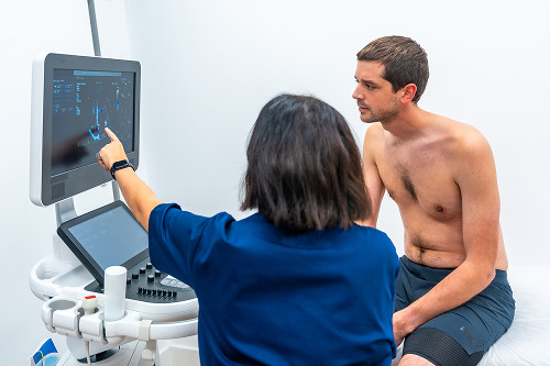
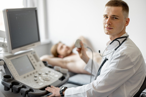
How is an echocardiogram performed?
An echocardiography service is a meticulously designed, calm, and comfortable process for the patient from start to finish. You can have an echocardiogram in Moscow at the K+31 clinic.
Echocardiography is a highly informative and completely painless diagnostic procedure performed in a comfortable environment for the patient. It follows a clear, well-established algorithm:
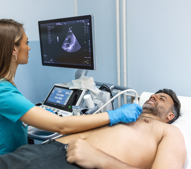
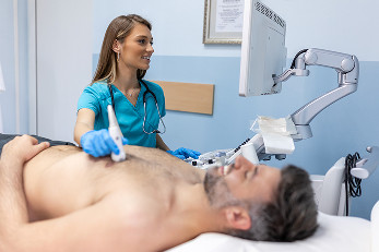
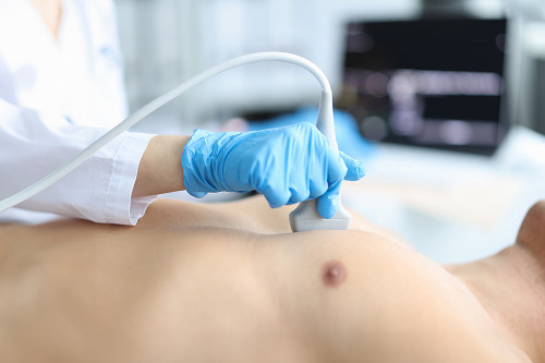
- Preparation and positioning The patient is invited into the ultrasound room and asked to lie down on the examination table. To obtain the best images, the examination is performed in the left lateral decubitus position. This slightly shifts the heart toward the anterior chest wall, exposing its structures for a more detailed examination
- Application of transducers and gel application The diagnostician applies a small amount of a special hypoallergenic gel to the skin over the heart. It is cool, but the sensation quickly passes and causes no discomfort. This gel eliminates the air gap between the transducer and the skin, ensuring ideal acoustic contact and high-definition imaging
- Scanning process The technician smoothly moves the ultrasound transducer across the chest. The patient is not required to do anything – simply lie still and briefly hold their breath at the doctor's request. This ensures static images are obtained and eliminates artifacts from lung movement. The scan itself is comfortable, with only a slight feeling of pressure. The entire procedure, depending on the complexity of the case, takes between 20 and 40 minutes
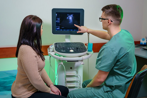
- Completion and results After the examination, any remaining gel can be easily removed with a disposable wipe. The patient will not need any recovery time – they can immediately return to their normal routine. The examination report, including a breakdown of all parameters and the doctor's conclusions, is provided immediately, within 15-20 minutes. This allows for prompt next steps – a consultation with a cardiologist to interpret the results and determine further treatment
General information
Preparation for Echocardiography
One of the significant advantages of this modern diagnostic method is the lack of complex and time-consuming preparation. The patient does not need to follow a diet, fast, or adjust their medication regimen, unless otherwise discussed with the attending physician in advance. However, to ensure the clinic visit is as comfortable and effective as possible, it is recommended to consider a few simple organizational points.
- Clothing. Since the examination will be performed on the chest, it is optimal to wear comfortable, non-restrictive clothing that can be easily removed or unbuttoned. Separate clothing (such as pants and a T-shirt) is ideal, as well as shoes that can be quickly and easily removed.
- Important Documents. To ensure a complete and informative medical examination, the diagnostician may require medical history information if the examination is not being performed for the first time. Be sure to bring: a doctor's referral (if available), stating the preliminary diagnosis and objectives of the examination; Previous ECG (electrocardiogram) results; previous cardiac ultrasound data (if performed) – this will allow the doctor to assess the dynamics of changes; outpatient records or hospital discharge summaries related to cardiovascular diseases.
- Emotional state. It is important to remain calm and not worry. The procedure itself is comfortable and not associated with discomfort. It is advisable to avoid intense physical activity and caffeinated beverages (strong tea, coffee, energy drinks) 2-3 hours before the clinic visit, as they can temporarily increase heart rate and affect some cardiac parameters.
Therefore, preparation means, first and foremost, sensibly organizing the visit. All these simple steps will help the doctor conduct the examination as thoroughly as possible, and the patient will receive comprehensive information about their health status without unnecessary stress and time expenditure.

Frequently Asked Questions
How long does the procedure take?
Is the procedure safe for children and the elderly?
Is any special preparation required for the examination?
When will the report be ready?
Will there be any discomfort during the examination?
Can I have an ECHO-CG without a doctor's referral at my own request?
ECHO-KG at the K+31 Clinic
The K+31 Clinic eliminates these disadvantages: our medical center is equipped with modern ultrasound equipment, including echocardiography. We prioritize patient health and conduct all examinations using the latest equipment, minimizing the risk of diagnostic error.
Our clinic also employs specialists with the highest level of ECHO-KG expertise. All of this allows us to accurately and reliably diagnose, prescribe the right treatment, and maintain your health.

Our doctors


This award is given to clinics with the highest ratings according to user ratings, a large number of requests from this site, and in the absence of critical violations.

This award is given to clinics with the highest ratings according to user ratings. It means that the place is known, loved, and definitely worth visiting.

The ProDoctors portal collected 500 thousand reviews, compiled a rating of doctors based on them and awarded the best. We are proud that our doctors are among those awarded.
Make an appointment at a convenient time on the nearest date
Price

Appointment to the doctor
Reviews


Our clinics

Application “Personal Account K+31”


What is echocardiography?
Echocardiography (ECHO) or cardiac ultrasound is a study of the heart using ultrasound that evaluates:
What is the difference between ECHO and cardiac ultrasound?
From a medical point of view, ECHO and Cardiac ultrasound is an absolutely identical examination, the same highly informative imaging method. The only difference lies in the accepted terminology.
Echocardiography is the full, official medical term. It is derived from the Greek words "ἠχώ" (echo), "καρδία" (heart), and "γράφω" (to write). The name accurately reflects the physical basis of the method—the recording of reflected ultrasound waves.
Cardiac ultrasound is a more simplified, colloquial term, understandable and familiar to a wide range of people. It is a synonym, emphasizing the technology used in diagnosis (ultrasound).