


About the disease
Anomalies of the development of the genital organs include congenital disorders of their anatomy due to incomplete organogenesis. In this case, there are changes in size, shape, proportions, symmetry, topography, and sometimes the complete absence of an organ.
Today it is possible to correct congenital pathology of the genitals using various types of surgical interventions.
Intrauterine formation of the genitals
The organs of the genitourinary system are formed from the genitourinary fold, which is formed as early as 2 weeks of intrauterine development. At 4-5 weeks, two female (Müllerian) ducts are formed from the crease, which then merge at 7-11 weeks to form the cervix and vagina, and at 13-14 weeks form the uterus and fallopian tubes.
The uterine cavity is initially represented by two uterine-vaginal cavities separated by a medial septum. By 16 weeks of intrauterine development, this septum gradually dissolves: initially, the bicuspid uterus takes a saddle shape, and then a normal, pear-shaped, single-cavity shape. At the age of 1 to 7 years, the size of the uterus does not change significantly. The cervix is not clearly differentiated at this age.
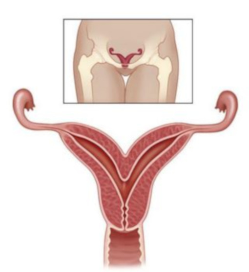
Bipedal and saddle-shaped uterus
In cases of incomplete processes of uterine formation by the time of birth, the uterus may remain either bicornal or have The uterus may remain either double-horned at birth or have some degree of cleavage: flattening of the fundus, weak divergence of the fundus into two horns. In all cases, the shape of the uterus in section resembles a saddle. Abnormalities of the uterus are often combined with malformations of the urinary system and a narrow pelvis.
Causes of impaired embryogenesis are various damaging factors that affect the fetus during pregnancy:
Malformations of the uterus are usually detected by chance. A woman may not be aware of a congenital pathology. A pronounced malformation of the uterine floor often results in primary infertility, which causes the patient to seek to a doctor. Standard gynecological examination for saddle-shaped and bicornic uterus is low-informative. In diagnosing Congenital uterine anomalies the decisive role belongs to instrumental studies - ultrasound pelvic organs, hysteroscopy, Hysterosalpingography, magnetic resonance imaging (MRI). When a malformation is detected, surgery is used to restore the normal uterine anatomy, surgical treatment is used. After correcting the birth defect, the chances of conceiving and carry a baby dramatically increase.
A slight saddle-shaped deformity does not prevent conception. But, nevertheless, during pregnancy can be detected: placental abnormalities (lateral or low location, placenta previa placenta previa, premature detachment), abnormal position of the fetus, premature birth, weakness or discoordination of labor. Anatomical and functional insufficiency of the uterus can provoke postpartum bleeding.

Appointment to the doctor
Lack of vagina
Due to the underdevelopment of the lower parts of the Müllerian passages, the vagina may be completely or partially absent (aplasia) - Mayer-Rokitansky-Küster-Hauser syndrome. The uterus and fallopian tubes also have abnormal structure. In addition, vaginal aplasia is often associated with abnormalities in the urinary system (kidneys, ureters, and the urinary tract) and the spine.
Despite the fact that the occurrence of this defect is due to a genetic predisposition, women with this pathology have a typical set of chromosomes (46, XX). They have well-formed external genitals, secondary sexual characteristics are developed, there are normal ovaries. The disease is characterized amenorrhea, sexual life becomes impossible. On examination, the complete absence or underdevelopment of vagina and often the uterus. For the final diagnosis, ultrasound of the pelvic organs and kidneys is used, which allows you to identify changes in the structure of the organs of the genitourinary system. MRI is used to determine the type of defect.
The treatment of vaginal aplasia is done surgically by laparoscopy. The vagina is created artificially from a skin flap, pelvic peritoneum, section of the sigmoid or rectum, as well as using alloplastic materials. Today, the first data from American scientists about artificially grown from the patient's own cells and transplanted vagina (Raya-Rivera A.M. et al., 2014). In case a woman has her own rudimentary vagina, it is possible to bougie it using a special device.
Infection of the vagina
Due to the inflammatory process during fetal development, atresia of the vagina may occur - complete or partial infection of a normally formed vagina. At the same time, the external genitalia, uterus, cervix, tubes and ovaries are developed and function correctly.
Due to atresia of the vagina after puberty, the outflow of menstrual blood from the uterus is disturbed, sexual life becomes impossible or difficult. Such patients periodically experience sharp pains in the lower abdomen, but external menstrual bleeding does not occur.
Congenital atresia of the vagina is often combined with the infection of the anus and / or underdevelopment of the organs of the urinary system. Complications of this pathology can be: the development of an ascending infection, peritonitis, sepsis, re-fusion of the walls of the vagina. Atresia of the vagina prevents the onset of pregnancy and the normal course of childbirth.
This defect is diagnosed during gynecological examination, vaginal probing, ultrasound and MRI of the pelvis. The treatment is exclusively operational - the restoration of a full-fledged vagina.

Septum of the vagina
If the intrauterine fusion of the Müllerian ducts is disrupted, a septum may form in the vagina. If it is located longitudinally, then more often it does not interfere with sexual activity and the onset of pregnancy, but in rare cases during childbirth can interfere with the progress of the presenting part of the child.
Transverse partitions, on the contrary, narrow the lumen of the vagina, which after puberty can cause the absence of menstruation. As a result, in a girl, the blood accumulated above the septum stretches the upper part of the vagina, fills the uterus (hematometra), fallopian tubes (hematosalpinx), enters the small pelvis, which provokes the occurrence of sharp pains in the lower abdomen.
The presence of a vaginal septum requires surgical intervention if it prevents outflow of menstrual blood or the birth of a fetus. In the latter case, its dissection is possible directly in childbirth.
Atresia of the hymen
Atresia is the complete absence of an opening in the hymen. Such a defect is manifested by amenorrhea, since due to an obstruction at the level of the hymen, blood cannot leave the vagina. It accumulates, stretching the walls of the vagina, then it can fill the cavity of the uterus and fallopian tubes, penetrate into the small pelvis in the same way as it happens in the presence of a transverse vaginal septum.
A girl with hymen atresia has no menses after puberty. At the same time, periodically pulling and cramping pains in the lower abdomen, weakness and dizziness due to the accumulation of blood in the vagina, uterus, fallopian tubes and small pelvis.
When examining the external genitalia, you can find a protruding hymen bluish color due to translucent blood. The final diagnosis is established on the basis of ultrasound of the pelvic organs, which allows you to detect blood accumulated in the genital cavities. With atresia of the hymen, surgical treatment is performed - a cruciform dissection of the hymen or partial excision.
At present, it is possible to eliminate almost any congenital anomaly of the genital organs, restore menstrual and reproductive function of women. The main thing is to turn to experienced specialists in a timely manner
Price
Make an appointment at a convenient time on the nearest date
Our doctors


This award is given to clinics with the highest ratings according to user ratings, a large number of requests from this site, and in the absence of critical violations.

This award is given to clinics with the highest ratings according to user ratings. It means that the place is known, loved, and definitely worth visiting.

The ProDoctors portal collected 500 thousand reviews, compiled a rating of doctors based on them and awarded the best. We are proud that our doctors are among those awarded.
Reviews


Our clinics

Application “Personal Account K+31”


