CT enterography

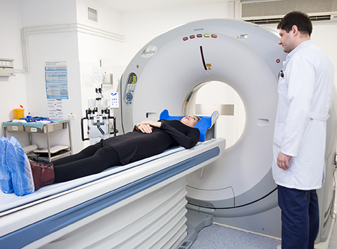
CT enterography or computed tomography of the intestine is a modern method of radiation diagnosis of diseases and pathologies of the small intestine. The essence of the procedure is to fill it with a neutral medium in order to straighten the lumen of the intestine and achieve the necessary contrast of the walls. If it is necessary to obtain high-contrast visualization of the mucous membrane and feeding vessels, then the diagnosis is carried out with intravenous bolus contrast enhancement.

Benefits of using CT enterography

CT enterography allows you to assess the condition of the walls, mucous membranes and feeding vessels in the intestine much better and more accurately than any other procedure. It allows you to obtain information about the pathologies not only of the small intestine, but also of other organs of the small pelvis, abdominal cavity and retroperitoneal space.

In the course of preliminary preparation for diagnosis, the patient's intestines are cleaned and stretched with liquid contents, which makes it possible to identify diseases and pathologies at an early stage, starting their treatment in a timely manner. During the procedure, the doctor can assess the condition of the mucous, muscular and serous membranes of the small intestine, the nature of the changes in which accurately indicates a particular disease. Polyethylene glycol preparations used in diagnostics are absolutely safe for human health.

But, like any other medical procedure, CT enterography of the small intestine has its own contraindications:

  • "Acute" belly.
  • Intestinal obstruction.
  • Allergy to individual components of the drug.

In the presence of such contraindications, the decision on the advisability of CT enterography of the small intestine is made by the doctor individually, depending on the patient's condition. Doctors should be informed in advance about recent infections and chronic diseases (diabetes, asthma, thyroid pathology, kidney failure).

Preparation for diagnostics

CT enterography is performed in a hospital setting. There is a misconception that before a CT scan of the small intestine, you need to sit on a special diet for 2-3 days. In fact, it is enough to abstain from food for 4-5 hours - and you can conduct a study. Drinking water in preparation for CT enterography is allowed.

Immediately before the procedure, it is necessary to drink several glasses of a solution that contains a contrast agent. In bolus contrast, contrast is injected intravenously into the patient using an automatic injector. Non-ionic iodine-containing preparations are used at the rate of 2 ml per 1 kg of the subject's weight.

Indications for CT enterography

This study is prescribed by a doctor for the following symptoms:

  • Indigestion.
  • Vitamins that enter the body with food are not absorbed.
  • Instability of the stool, bloating and pain in the abdomen.
  • Preparation for surgery.
  • Monitoring the effectiveness of therapy.
  • Chronic diseases that require regular diagnosis of the condition of the gastrointestinal tract.
  • Inflammatory lesions of the small intestine, particularly in Crohn's disease.
  • Suspicion of a tumor of the small intestine.
  • Malabsorption syndrome (celiac disease).
  • Occult gastrointestinal bleeding.

How is CT enterography performed?

CT enterography is performed using a tomograph by transilluminating the examined area with x-rays. The radiation dose is minimal, so the procedure is absolutely safe. Passing through the body, X-rays make it possible to obtain layer-by-layer detailed images and a three-dimensional model of the organ under study. Signals are captured by touch sensors, and all data is processed by a computer.

CT enterography takes an average of 40 minutes. Rehabilitation after diagnosis is not required, the contrast agent will leave the body naturally.

CT enterography of the small intestine is absolutely painless, unlike colonoscopy and other instrumental methods of examination. During the procedure, the patient will not even feel discomfort. The only exception is that when a contrast agent is injected, a metallic taste may be felt in the mouth, but it quickly passes.

You can undergo CT enterography as prescribed by a doctor at the K+31 clinic. Among the advantages of contacting us:

  • Our medical center uses modern computed tomography equipment that meets international standards.
  • The result of CT enterography (images and a detailed conclusion of the diagnostician) in electronic form will be ready within 30 minutes after the completion of the diagnosis.
  • Examination of the small intestine is carried out by appointment, no queues and affordable prices for patients.
  • Other research methods are available: MRI of the intestine, colonoscopy, EGD of the stomach and other studies of radiation diagnostics.

Service record