How?
Regular digital mammography is the only method for early diagnosis of breast cancer with proven effectiveness. It is a reliable, fast, inexpensive diagnostic method to rule out health problems before symptoms appear.
Authoritative medical communities from different countries are unanimous in the opinion that mammography is optimal as a screening (that is, preliminary) method for examining women.
If necessary, after mammography, the patient is referred for other diagnostic procedures.
It must be remembered that in a group of women at high risk of developing breast cancer (identified mutations in the BRCA1 or BRCA2, p53, PTEN, CDH-1, CHEK2 genes, a burdened family history, radiation therapy for lymphogranulomatosis), the examination begins with an annual magnetic resonance imaging contrast-enhanced tomography at age 25.
When?
If nothing bothers you, the examination must be taken from the age of 40. One of the largest international studies (link to article) showed that performing mammography once a year from the age of 40 reduces mortality from breast cancer by 50%.
If you have changes in your breasts (skin color, shape, nipple discharge, pain).
If there is a genetic predisposition (mutations in the BRCA1 or BRCA2, p53, PTEN, CDH-1, CHEK2 genes have been identified), mammography must be performed starting at the age of 35 in addition to the annual magnetic resonance imaging.
What for?
The main purpose of the annual examination is to detect breast cancer at stages 0 and I.
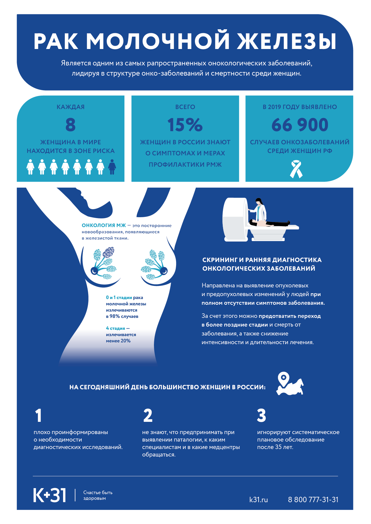
According to the World Health Organization, every 8 women in the world develop breast cancer. However, at an early stage (0 and I) -99% of women live without a relapse of the disease, 98% of women undergo organ-preserving surgery, which allows them to preserve the aesthetic side of life.