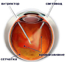
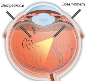
Vitrectomy Surgery Technique
During a vitrectomy, the surgeon inserts the finest instruments into the eye, cuts off the micro-pieces of the vitreous, and aspirates it. After removal of the vitreous, the surgeon can begin to treat the retina with a laser (laser endocoagulation), cut or remove fibrous or scar tissue from the retina, straighten areas where the retina exfoliates, remove traction and remove holes in the retina or macula.
At the end of the operation, in order to replace the vitreous body and restore normal pressure in the eye, a special balanced saline solution, silicone oil or gas is injected.
Vitrectomy is always performed by a surgeon who has special training in treating retinal problems. Doctors of the Ophthalmology Department. Eye microsurgery K+31 are highly qualified specialists in the field of microsurgical operations.
What to expect after surgery
Vitrectomy can be performed on an outpatient basis, but, in some cases, may require hospitalization. The operation lasts from 1 to 3 hours and is performed under local or general anesthesia, depending on the recommendations of the surgeon and anesthetist.
After the operation at home for some time, you will have to observe a certain position of the head. The surgeon will tell you which one, depending on the pathology and on what the vitreous body was replaced with.
How well vitrectomy helps
Vitrectomy significantly improves visual acuity in patients with vitreous hemorrhage that does not resolve. May reduce the risk of serious complications of chronic bleeding into the vitreous body and the growth of abnormal blood vessels.
Vitrectomy, at times, is the only method for eliminating retinal detachment and severe proliferative vitreoretinopathy.
An operation can restore some vision that is lost as a result of tractional retinal detachment and can help prevent further detachment. But the results are usually better when the detachment does not affect the center of the retina (macula) and the central vision is maintained.
Vitrectomy Risks
Vitrectomy can cause increased pressure inside the eye, especially in people who have glaucoma.
There are several other complications associated with vitrectomy. These include:
- Ongoing bleeding.
- Relapse of retinal detachment.
- Corneal edema.
- Infection inside the eye (endophthalmitis).
Surgical treatment of the vitreous body and retina
K+31 uses the latest advances in vitreous and retinal surgery. Doctors are constantly improving their skills in Europe and the USA.
Operations are carried out on the most modern surgical equipment. All surgical techniques are minimally invasive, less traumatic, for the most part seamless and painless.
Control over the general condition of the patient during surgery is carried out by experienced anesthetists.
All risks are minimized through the use of expensive disposable supplies from world famous manufacturers (Bausch & Lomb, Alcon, Dorc, BD, Synergetics, Geuder).
- K+31 is a premium quality of medical care at average market prices.
- In K+31, you can solve several problems in one visit.
- K+31 - these are extra-class specialists who receive 42 medical specialties and perform complex operations / solve complex medical problems. The clinic has 2 academics and 2 corresponding members of the RAMS, 19 professors, 19 doctors of medical sciences, 27 candidates of medical sciences.
- K+31 is an ultramodern equipment that helps to solve the most complex medical problems, allows the most advanced diagnostic equipment (CT, MRI, endoscopy, x-ray) and hospital equipment.