Retinal dystrophic changes

Retinal dystrophy is a condition accompanied by a violation of its nutrition. Metabolic processes are disrupted at the cell level, then functional and structural changes are noted.

Diseases and conditions leading to retinal dystrophy:

  • Diabetes.
  • Arterial hypertension.
  • High myopia.
  • Common atherosclerosis.
  • Traumatic brain injury.
  • Eye damage.
  • Consequences of infectious and viral diseases.

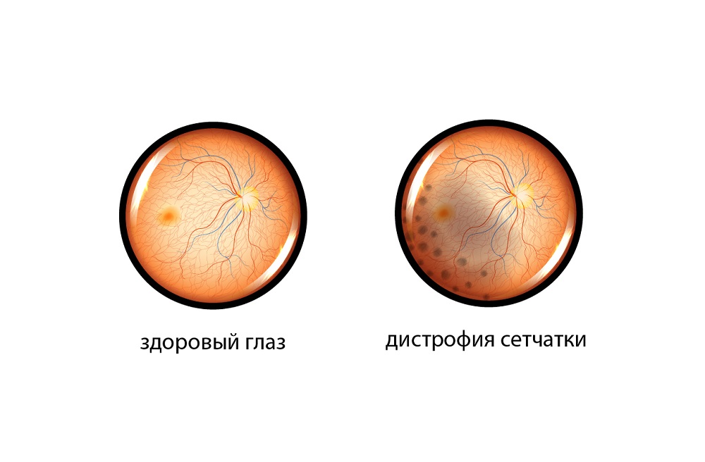
Allocate central retinal dystrophy, in which changes develop in the macula - the exit point of the optic nerve. This area is responsible for visual acuity and perception of small objects. In the early stages, the disease may not manifest itself in any way. Later, vision deteriorates, dark spots appear in front of the eyes and difficulties in focusing on small details.

The second type is peripheral dystrophy. The foci of pathology are located outside the macula, which is why they do not manifest themselves in any way. Peripheral dystrophy is dangerous with retinal detachment, therefore, laser coagulation is indicated.

An ophthalmologist deals with the diagnosis of pathology. In the K+31 clinic, it is possible to undergo examination and treatment using modern equipment that allows you to accurately identify and successfully eliminate degenerative changes in the retina.

Indications

Conditions in which the operation is required: retinal detachment or rupture, lattice thinning, dystrophy of the "snail trace" type, the presence of blood clots in the vessels of the eye, diabetic changes, macular edema.

Contraindications

The operation is not performed in the presence of hemorrhage in the eye cavity, cataracts, formation of gross gliosis (gliosis is an overgrowth of connective tissue that can grow into the vitreous body, disrupting the functioning of the organ of vision). Laser correction can be performed after elimination of the pathology.

Retinal dystrophy treatment

Timely start of treatment helps to stop the pathological process and maintain visual acuity. The types of impact in this case are as follows:

  1. Medication. Includes drops that improve retinal metabolism. Therapy is aimed at maintaining the condition, preventing its deterioration.
  2. Physiotherapy. Electrophoresis is used, as well as exposure to a magnet, laser.
  3. Surgical. The main method of treatment that eliminates the pathology completely. The recommended type of exposure is laser coagulation of the retina.

The essence of the procedure: the doctor acts with a laser beam on the weak areas of the retina, thereby strengthening them and preventing further thinning.

Retinal laser coagulation

The operation is performed on an outpatient basis. The patient sits down in front of the slit lamp with the chin on a special holder. The doctor conducts an examination through a laser correction machine, while an enlarged image of the retina with various parameters and characteristics is displayed on the screen.

Operation progress:

  1. The ophthalmologist puts a lens on the eye to prevent involuntary reflex movements, asks the patient to look straight.
  2. Directs the laser beam to problem areas. At the same time, the patient sees red dots, flashes of light, hears the sound signal of the equipment.
  3. The patient looks up, at this moment the lens is removed.

The duration of the procedure is from 5 to 15 minutes. Due to the use of local anesthesia, laser treatment is well tolerated by patients. Upon completion, drops are instilled into the eyes.

Laser coagulation of the retina can be performed for pregnant women up to 36 weeks of gestation. At a later date, the procedure is not performed, since the adhesions that can withstand the load during childbirth will not have time to form. Previously transferred laser coagulation in most cases is not a contraindication to natural delivery.

Preoperative preparation

Two weeks before the intervention, you cannot use hard type contact lenses, two days - soft type. No other preparatory procedures are required from the patient. As diagnostic manipulations 3-7 days before the expected date of the operation, a repeated examination by an ophthalmologist may be prescribed to assess the dynamics of pathological changes.

Immediately before the operation, the doctor instructs the patient on how to behave during the procedure:

  • Listen to what the doctor says and follow through.
  • Do not make sudden movements with your eyes.
  • Don't move your head.

How to behave after surgery

To speed up healing and prevent complications, the doctor prescribes anti-inflammatory and healing drops.

What not to do within two weeks after surgical treatment of retinal dystrophic changes:

  • Lift objects heavier than 3 kg.
  • Instill medications in the eyes without a doctor's prescription.
  • Drink alcoholic beverages.

Forecast and results of surgery

Timely manipulation allows you to maintain visual acuity and prevent retinal detachment. As with any operation, there are risks with laser coagulation, but they are small compared to the possible consequences of pathology.