Artificial intelligence helps accurate diagnosis at K+31 clinic
K+31 Clinic was one of the first in Moscow to implement artificial intelligence for processing CT scans of the chest organs. This helps to make a preliminary diagnosis as quickly as possible, saving time on analyzing images and preparing reports.
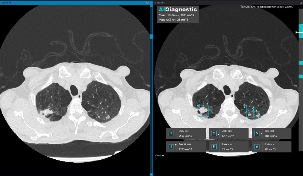
Here's how it works: CT scans are viewed and evaluated not only by a doctor, but also by AI: it finds pathological foci, measures them, marks them and calculates the volume.
“As a result, this saves the doctor’s time – he only needs to review the study processed by AI and draw up a protocol based on the already calculated indicators,” explains the doctor of the K+31 diagnostic center.
Artificial intelligence effectively identifies potentially malignant lesions in the lungs, calculates the volume of hydrothorax (pathological accumulation of fluid in the pleural cavity), evaluates signs of aortic aneurysm and pulmonary trunk expansion.
For patients, the advantage of such diagnostics is that the use of AI reduces the human factor: the risks of errors and omissions are reduced. In essence, the patient receives a double opinion – the image is assessed by both the doctor and the AI.
The process is arranged as follows: the study from the diagnostic device of the CT machine goes to a special service (AI Diagnostic), which analyzes the image and returns the result to the workstation of the radiologist. Image processing takes about 1 minute. All studies are pre-anonymized and all personal data is under reliable protection.
“By using AI for radiation diagnostics, we at K+31 support modern trends in healthcare,” the doctor explains. “It is important for patients to understand that artificial intelligence is an assistant, not a replacement for a doctor. Its task is to help the doctor not to miss pathologies, to notice negative or positive dynamics in time in order to prescribe the most effective treatment.”


