Поиск
ENDOCRINOLOGIST APPOINTMENT
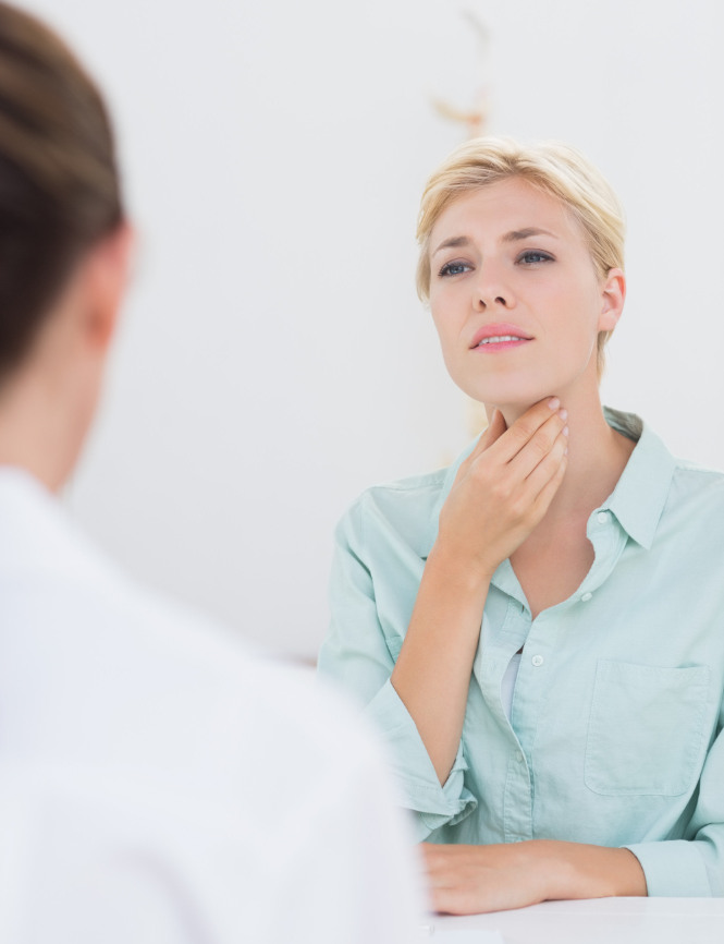
When to see an endocrinologist? The endocrine system is one of the key regulators of the processes occurring in the body. Many will be surprised to learn that the endocrine system regulates not only weight and reproductive capacity, but also blood pressure ...
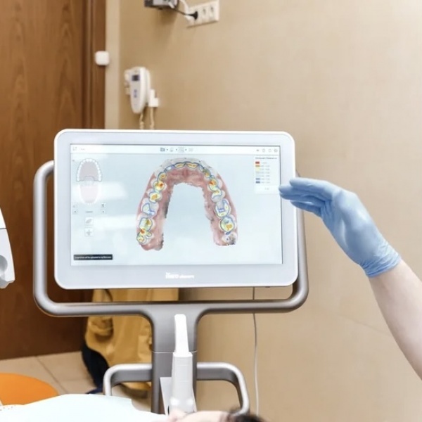
3D SCANNING
The dentistry department K+31 Petrovskie Vorota has new opportunities for the patient - the iTero Element intraoral 3D scanner. This is modern equipment of high precision, thanks to which it is possible to predict the results of treatment and do without ...
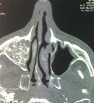
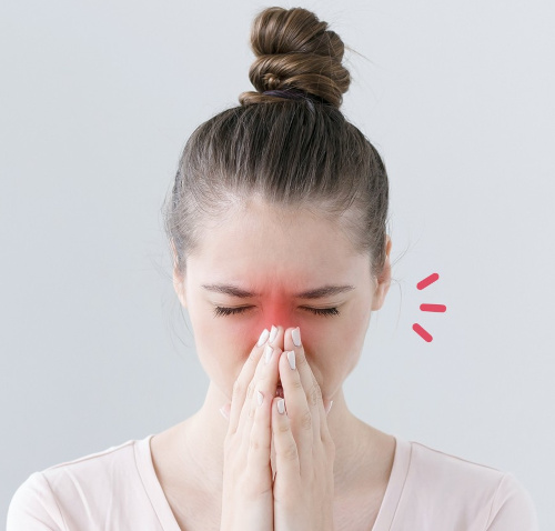
FOREIGN BODIES OF THE NASAL CAVITY AND SINUSES
Foreign bodies of the nasal cavity and sinuses are very diverse, at times, the history of their entry is quite fantastic. "Champions" in this regard are children who stick everything in their noses. If the foreign object is small and smooth, blowing out ...
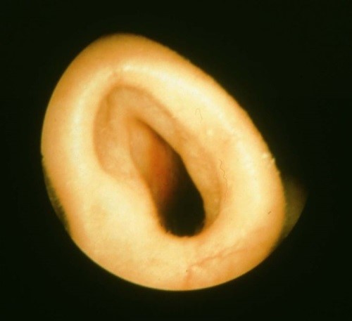
INSUFFICIENT NASAL VALVE
The valve of the nose is the narrowest point in the upper airway, which ensures the "swirling" of the air flow, which, moving turbulently, travels the greatest distance in the nasal cavity. During this movement, the air we breathe is humidified, warmed or ...
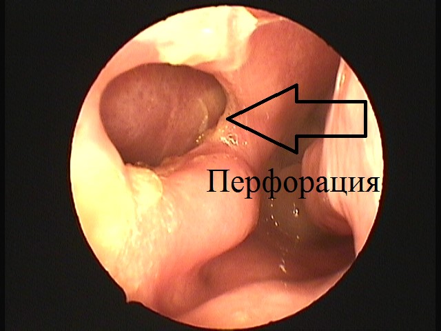
TREATMENT OF PERFORATION OF THE NASAL SEPTUM
Perforation of the nasal septum develops: Due to surgery on the septum of the nose. As a result of injury. Due to the uncontrolled use of vasoconstrictor drops. After using drugs. Due to the wrong toilet of the nasal cavity. Due to the development of ...
SILENT SINUS SYNDROME TREATMENT
Silent sinus syndrome (SMS) is a rather rare disease. In fact, this disease is a deformation of the walls of the maxillary sinus caused by various reasons, as a result of which the sinus is pressed inward. Such deformation leads to a change in the shape of ...
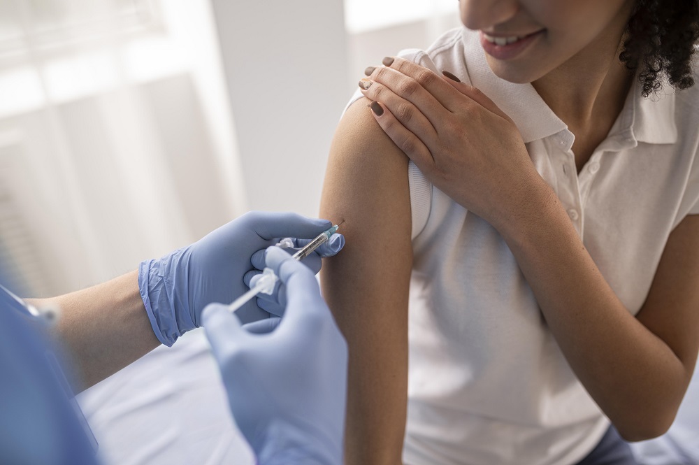
VACCINATION AGAINST TYPHOID FEVER
Why is vaccination required? Typhoid fever is a very dangerous bacterial infectious disease. It affects up to twenty million people in the world every year. This disease is most common among adolescents and children. If an advanced disease is left ...


