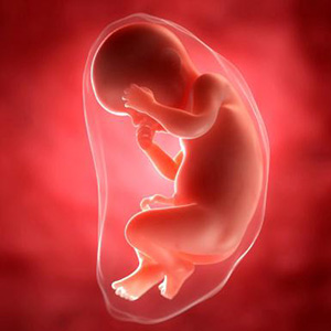
20 days after successful conception, serious changes occur in the woman's body associated with the intrauterine development of the fetus. After implantation into the uterine wall, the embryonic tissue begins to secrete chorionic gonadotropin (hCG), which leads to serious hormonal changes. After 2.5 weeks after conception, the objective signs of pregnancy become aggravated - toxicosis, a change in taste, an increase in basal and general temperature.
Feelings of a mother
 Within 20 days from conception, a colossal hormonal change occurs in the female body, associated with an increase in the level of estradiol, progesterone and hCG in the blood. Physiological changes are accompanied by a number of symptoms, which include:
- drowsiness;
- rapid fatigue;
- dizziness;
- mood swings;
- morning sickness;
- breast tenderness;
- swelling of the mammary glands;
- increased basal temperature;
- frequent constipation;
- violation of sleep and wakefulness.
During the final stage of the IVF procedure, three- or five-day-old embryos are used for implantation into the uterine cavity. According to clinical data, the latter are more likely to take root, which is associated with their maximum readiness for implantation into the endometrium. Discomfort in the lower abdomen and nausea are characteristic sensations in 20 DPPs of five days. To determine the features of the course of pregnancy during this period, the first ultrasound examination is performed.
What does an ultrasound examination show?
Ultrasound in 20 DPP is performed in order to detect the site of implantation of the ovum, which makes it possible to exclude the likelihood of developing an ectopic pregnancy. The examination is carried out using a transabdominal or transvaginal ultrasound sensor, which provides comprehensive information about the position of the fetus and the features of its development in a given embryonic period.
 Half an hour before the procedure, it is recommended to drink at least 1 liter of liquid, since the transabdominal examination is carried out under the condition of maximum filling of the bladder. With the help of ultrasound, the embryologist will be able to determine:
- localization of the ovum;
- the state of the endometrium and uterine mucosa;
- the size of the fetus and its age;
- physiological changes in the embryo.
Each embryonic period corresponds to certain features in the development of the fetus. Ultrasound examination allows you to determine the condition of the embryo and identify abnormalities if they occur.
On the 20th day after conception, on an ultrasound image, the embryo looks like a small elongated cylinder with embryonic limb development. On the monitor, you can see a small head and tail, which eventually develops into the limb.
Features of embryonic development after IVF
 In the first 20 days from conception, the embryo undergoes serious changes, ranging from gastrulation to organogenesis. During the embryonic period, which begins from the moment of fertilization until the 10th week of the obstetric period, the embryo grows rapidly in size.
At 2 and 3 weeks of gestation, the embryo does not resemble a baby and only at the stage of organogenesis gradually acquires characteristic outlines and structure. In the last week of embryonic term, the gill arches and tail disappear in the fetus.
Embryos at 20 days after conception
- On the ninth day after implantation into the uterine wall, the process of cell division of the embryo continues, as a result of which it is transformed from a single-layer blastosphere into a gastrula with several embryonic petals;
- On days 20-21, the embryo enters the neurula stage, which is characterized by the formation of a neural plate;
- At the beginning of the 4th week of gestation, the process of formation of the placenta begins. Chorion and amnion arise from the ectoderm and trophoblast, in which amniotic fluid subsequently begins to accumulate.
Chorion is a villous formation that produces hCG. With the participation of the mesoderm, a fetal place is formed from it, in which the fetus continues its further development. By the end of the third week of gestation, the fetus reaches a length of only 4-5 mm and looks more like an oblong cylinder.
Risks in the early stages of pregnancy
 According to medical data, the period between 20 and 28 days of gestation is the most dangerous for the expectant mother. It is during this period that the risk of spontaneous abortion and pregnancy fading increases. During IVF, the body is adjusted to bear the fetus artificially with the help of supportive hormonal therapy. Therefore, even the slightest fluctuations in the hormonal background can lead to fetal freezing.
In some cases, an ultrasound scan of 20 DPP shows that the embryo has attached to the wall of the fallopian tube, and not to the uterine cavity. In this case, an ectopic pregnancy is diagnosed, which must be terminated. To preserve the fetus and prevent complications, several important rules must be followed:
- Within 2-3 months after successful conception, you need to be observed in the clinic where the IVF procedure was performed;
- During the first month after conception, it is necessary to strictly control the content of progesterone in the blood;
- Vitamin-mineral complexes and hormonal preparations prescribed by a specialist should be taken, which maintain a normal hormonal background;
- Before the first ultrasound, it is advisable to abandon sexual intercourse in order to avoid surges in the hormonal background;
- It is necessary to avoid excessive physical exertion and stressful situations, which can affect the psycho-emotional state and provoke an increase in the tone of the uterus.
Conclusion
On the 20th DPP, the size of the embryo ranges from 4 to 5 mm. To exclude an ectopic pregnancy, a specialist often prescribes a first ultrasound examination. With a successful IVF, patients often have multiple pregnancies (usually twins), but in most cases, one of the embryos soon stops developing. On an ultrasound image, the embryo is not much like a baby. The characteristic outlines appear only at the final stage of embryonic development, which corresponds to the 10th week of the obstetric period.
